Quantcast
Channel: Microscope World Blog
Viewing all 422 articles
Browse latest View live

Mouse Embryo under Fuorescence Microscope


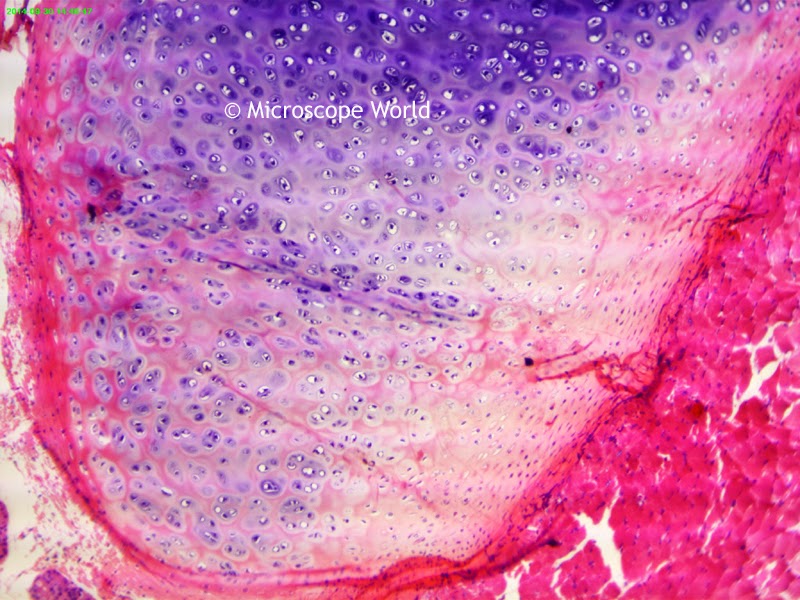
Bone and Cartilage Under the Microscope

$
0
0
Bones are rigid organs that support and protect various organs of the body, produce red and white blood cells and store minerals. Osseous tissue (sometimes referred to as bone tissue) gives bone its rigidity and coral-like internal three-dimensional structure. Other types of tissue found in bones include bone marrow, endosteum, periosteum, nerves, blood vessels and cartilage.

When a child is born he or she has over 270 bones in the body. Many of these fuse together as the infant grows and an adult human has 206 bones in the body. The largest bone in the human body is the femur in the leg, and the smallest is the stapes, a small bone found in the inner ear.

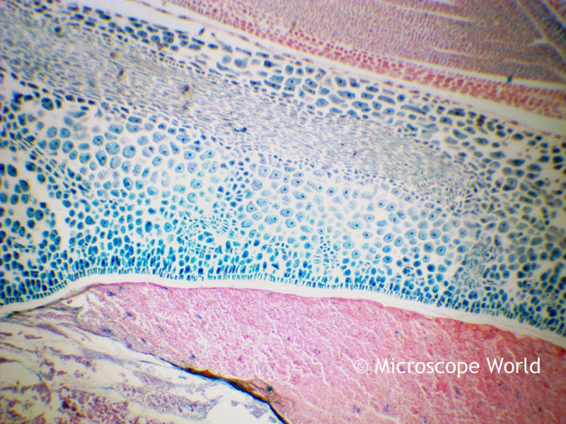
Bone and cartilage captured under a biological microscope at 40x magnification.
Bone and cartilage captured under the microscope at 40x magnification.
Cartilage is a flexible connective tissue that is not as hard as bone, but is stiffer and less flexible than muscle. Cartilage is found in the joints between bones, the rib cage, and even in the ear and nose. Unlike other connective tissue, cartilage does not contain blood vessels.

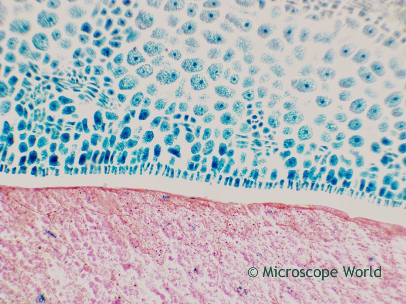
Bone and cartialge captured under a biological microscope at 100x magnification.
Bone and cartilage captured at 100x under a biological microscope.
Bone and cartilage prepared microscope slide can be purchased as part of the Histology Prepared Slide Kit.

Images of bone and cartilage were captured with the Richter Optica U2 biological microscope using a microscope digital camera.

Biological microscope image captured at 400x magnification.
Bone and cartilage captured at 400x magnification under a biology microscope.

House Fly Under Microscope!

$
0
0
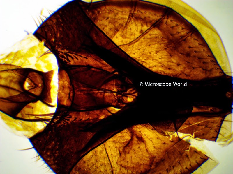
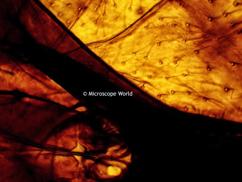
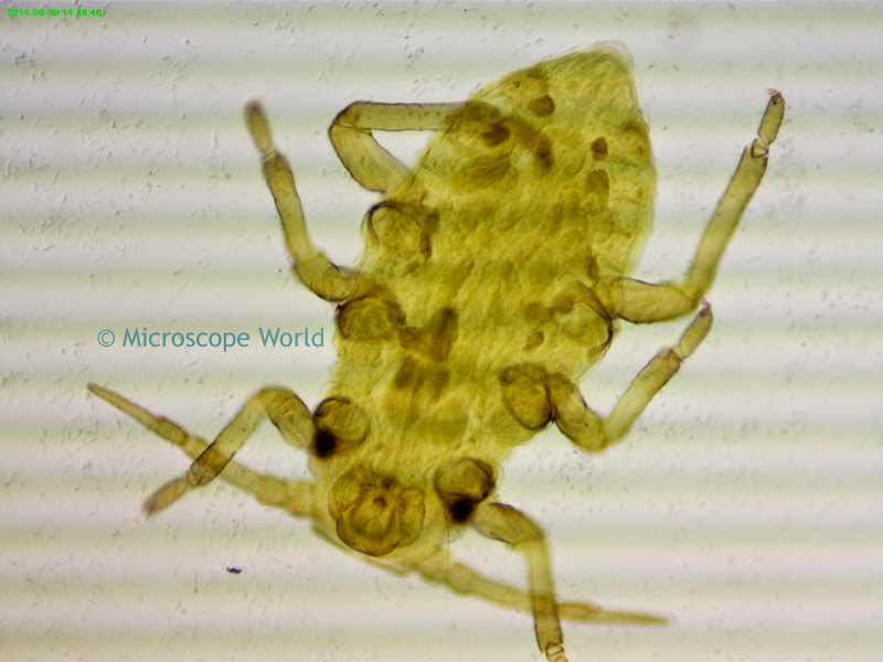
The common house fly is pesky and annoying. When viewed under a microscope it can be fascinating. These house fly images were captured by using the prepared slide from the Zoology, Entomology and Insect Prepared Slide Kit and placing it under the Richter Optica HS-1D digital high school biology microscope. The HS-1D digital microscope includes a 3 megapixel camera.

House fly under microscope at 40x magnification.
House fly captured under the microscope at 40x magnification.
House fly under microscope at 100x
House fly captured under the microscope at 100x magnification.
House fly under high school digital microscope at 400x magnification.
House fly captured under microscope at 400x magnification.

Student Science Project - Blue Water!

$
0
0
This is a fun kids student science project where you will watch a glass of clear water turn dark blue almost instantly! Gather some adult supervision, some safety goggles and wear old clothes (iodine can stain!)

Items needed:
  • 3 Clear glass cups
  • 1000mg of Vitamin C
  • Tincture of Iodine (2%)
  • Hydrogen Peroxide (3%)
  • Liquid laundry starch or 1/2 teaspoon corn or potato starch
  • Goggles for safety!
  • Measuring spoons & cups
  • Your adult for help & safety!
Science Project:
  1. Put on the goggles and mash your Vitamin C in a plastic bag, until it is a fine powder. Put all the powder in a glass with 2 ounces (60ml) of warm water. Stir it up. Label the glass LIQUID #1.
  2. Put 1 teaspoon (5ml) of LIQUID #1 into a new glass and add 2 oz (60ml) of warm water plus 1 teaspoon (5ml) of iodine. Notice the brown iodine turns clear! Label this glass LIQUID #2.
  3. In the third glass, place 2 ounces of warm water, 1 Tablespoon (15ml) hydrogen peroxide and 1/2 teaspoon (2.5ml) of the liquid starch. Label this glass LIQUID #3.
  4. Now for the fun! Pour all of LIQUID #2 into LIQUID #3 and then pour the liquid back and forth between the two cups several times to mix them well. Set the cup down on the table. Within a few seconds to several minutes your water should suddenly turn dark blue!

So What Exactly Happened?

You observed a chemical reaction called a clock reaction. It is called a clock reaction because if you try the experiment again, you can actually change the amount of time it takes for the liquid to change colors. The starch is trying to turn the iodine blue, and the Vitamin C is keeping it from turning blue. It is the moment when the Vitamin C loses the battle with the iodine, that the water instantly turns blue.

Try these variations:
  • Use different temperatures of water for your experiment and record the time difference for the water to turn blue. What makes the water turn blue faster - really hot or lukewarm water?
  • If you add more or less Vitamin C to LIQUID #1 does the water turn blue faster or slower?
Observation:
  • Once you have performed the experiment several times, place some liquid starch on a blank glass microscope slide and view it under the microscope
  • Place some of your blue water on a depression slide with a cover slip and view it under the microscope. How does the blue water look different from LIQUID #1?
  • Record your observations and share them with your class or science teacher.
  • Clean up the liquid by pouring it down the drain and washing all glasses and hands thoroughly.
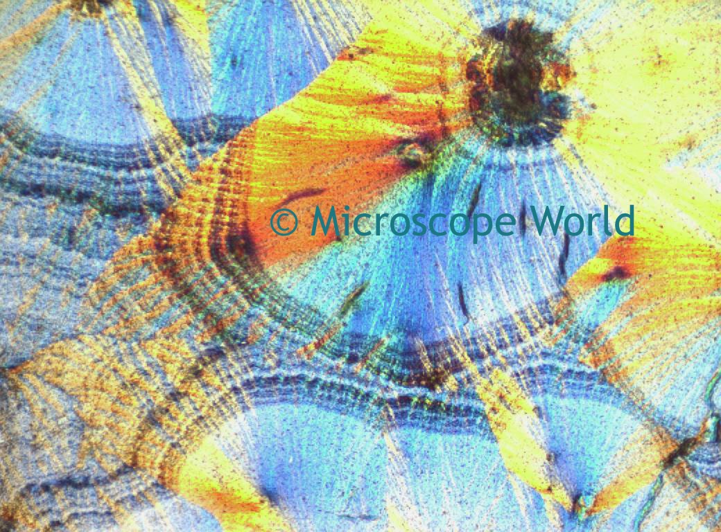
The image below of vitamin C was captured using a polarizing microscope. How does your vitamin C look different when viewing it under a student biological microscope?

Vitamin C captured under a polarizing microscope
Vitamin C under a polarizing microscope.

Gallbladder under the Microscope

$
0
0
The gallbladder is a small organ in vertebrates where bile is stored before being released into the small intestine. Humans can actually live without a gallbladder.


Under the microscope, layers of the gallbladder wall can be viewed. The gallbladder walls' innermost surface is lined with a single layer of columnar cells. The primary function of the gallbladder is to store bile, which is produced by the liver.

Gallbladder captured under a biological microscope at 40x
Gallbladder under the microscope at 40x.
These images were captured using the gallbladder microscope prepared slide. The images were captured with the Richter Optica U1 biological microscope and a microscope digital camera.

Gallbladder under the microscope at 100x magnification.
Gallbladder captured under the microscope at 100x magnification.
Microscope image of gallbladder captured at 400x magnification.
Gallbladder under the microscope at 400x.


Why Use a Stereo Microscope Auxiliary Lens?

$
0
0
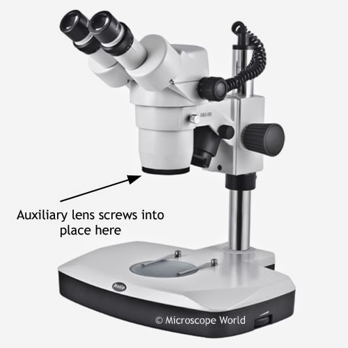
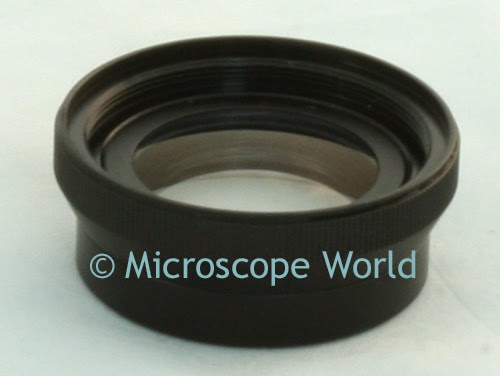
A stereo microscope auxiliary lens screws onto the base of the body of the stereo microscope.

Stereo microscope auxiliary lens from microscope world
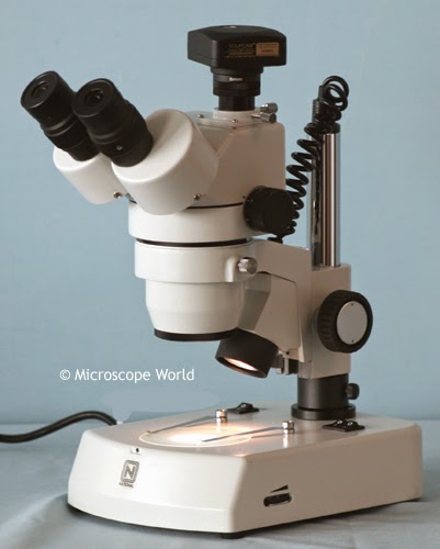
Stereo Microscope showing where Auxiliary Lens attaches.

There are several reasons why you might want to use a stereo microscope auxiliary lens. Sometimes using an auxiliary lens is the best way to increase or decrease magnification, especially if you are using a microscope camera. Microscope magnification can be changed by using different eyepieces such as 5x or 15x eyepieces. However, if you have a camera mounted on the trinocular port of your microscope, changing the eyepieces will not change the magnification that the camera sees. Adding an auxiliary lens will change the magnification you see both in the eyepieces as well as through the camera.

Stereo Microscope Auxiliary Lens from Microscope World
Stereo Microscope Auxiliary Lens

Stereo microscope auxiliary lenses are also used to change the working distance of the stereo microscope. Working distance is the amount of required distance between the top of your object and the bottom of the microscope lens in order for the sample to be in focus. Increased working distance is often desired when soldering, assembling printed circuit boards, or performing dental lab work.

Retina under the Microscope

$
0
0
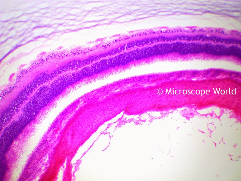
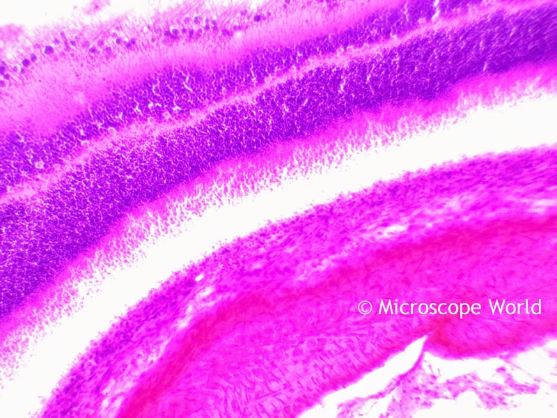
The retina is a light-sensitive layer of tissue that lines the inner surface of the eye. The optics of the eye create an image of the visual world on the retina, and it plays a very similar function as the film in a camera.

The images below of the retina were captured using a biological microscope and a 5 mega pixel microscope digital camera.

Retina under microscope captured at 40x magnification at Microscope World
Retina captured at 40x under the microscope.
 The retina microscope prepared slide is available in the Histology Microscope Prepared Slide Kit.

Retina captured under the microscope at 100x magnification at Microscope World
Retina captured at 100x under the microscope.

Retina captured at Microscope World at 400x
Retina captured at 400x magnification under the microscope.

Microscope Working Distance

$
0
0
Microscope working distance is the amount of distance required between the objective lens and the top of the object in order for the sample to appear in focus. This rule applies to both biological microscopes and stereo microscopes.

Typically the larger the magnification of a high power objective lens, or stereo microscope auxiliary objective lens, the shorter the working distance. As magnification increases, working distance decreases. This sometimes necessitates the use of a long working distance objective lens. Long working distance (LWD) objective lenses were created to allow greater working distance while using higher magnification.

High power objective lenses often have the working distance noted on the microscope objective, as shown above.

Sarcina under the Microscope

$
0
0
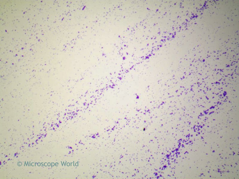
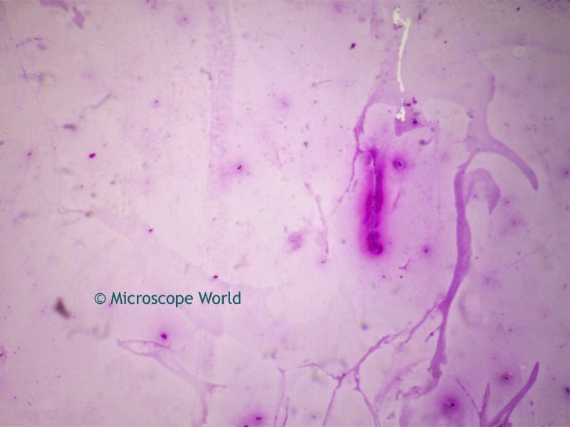
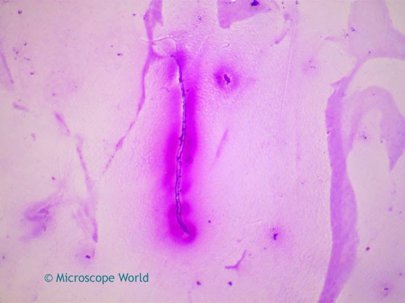
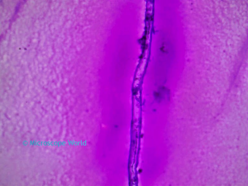
Sarcina is a genus of bacteria found most commonly in organic fluids in the stomach. The Sarcina prepared microscope slide is part of the Bacteriology Prepared Microscope Slide Kit.

Sarcina captured under the microscope at 40x magnification.
Sarcina captured under a biological microscope at 100x magnification.
Sarcina captured under the microscope at 100x magnification.
Sarcina captured under a biological microscope at 400x magnification.
All microscopy images were captured using the Richter Optica U2 biological microscope and a microscope camera with software.

Funghi Spores under the Microscope

$
0
0
A spore is a unit of asexual reproduction, often a part of the survival process. Spores can typically survive in unfavorable conditions and help form part of the life cycle of many plants including funghi. Spores are typically unicellular.

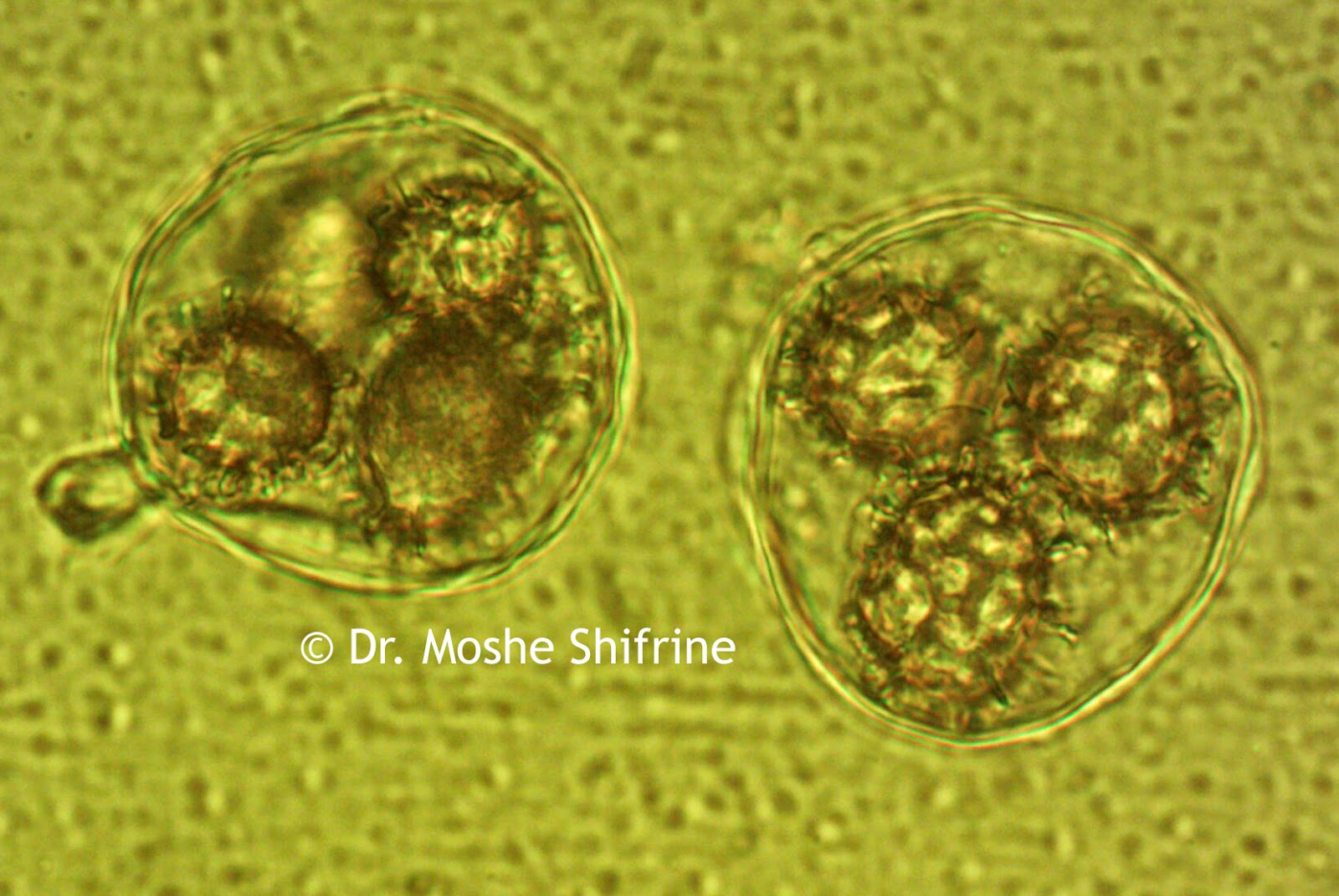
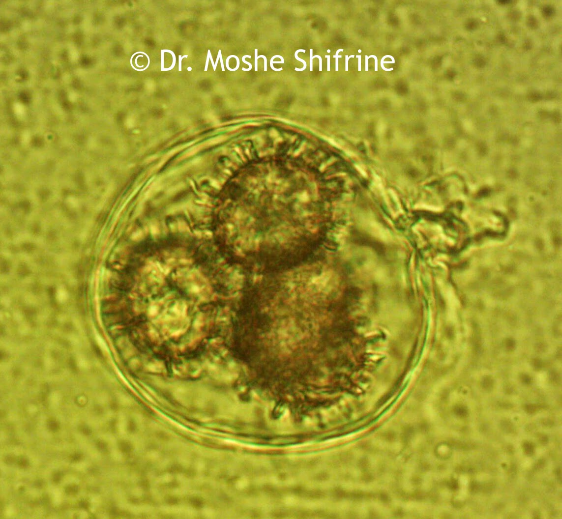
These funghi spore images were captured by Dr. Moshe Shifrine using the U2LCD digital tablet lab microscope. Dr. Shifrine is a retired Professor of Immunology (University of California at Davis) with a Ph.D. in microbiology specializing in mycology, the study of fungi or mushrooms.

Funghi spore captured under a biological microscope at 1000x magnification.
Funghi Spore under the microscope at 1000x magnification.

Funghi spore, 1000x, under biological microscope.
Funghi Spore captured under the microscope at 1000x magnification.
Funghi spore 1000x magnification.
Funghi spore under the microscope at 1000x.

Minerals Under the Microscope

$
0
0
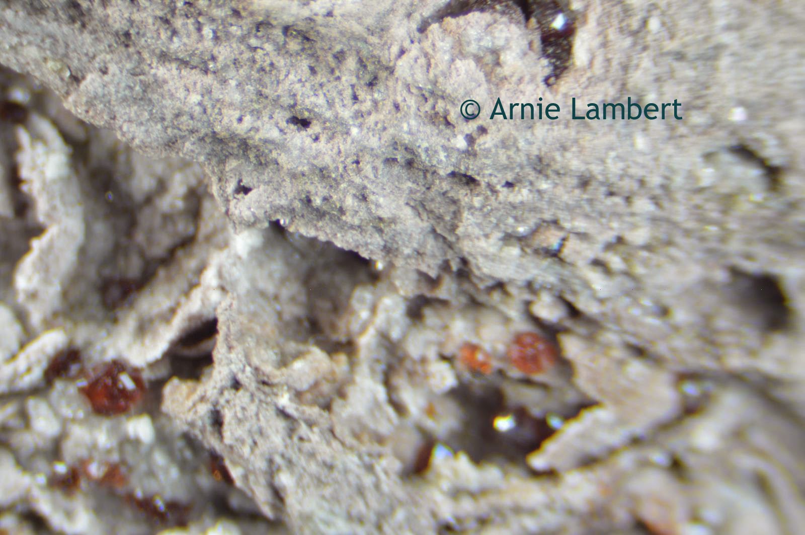
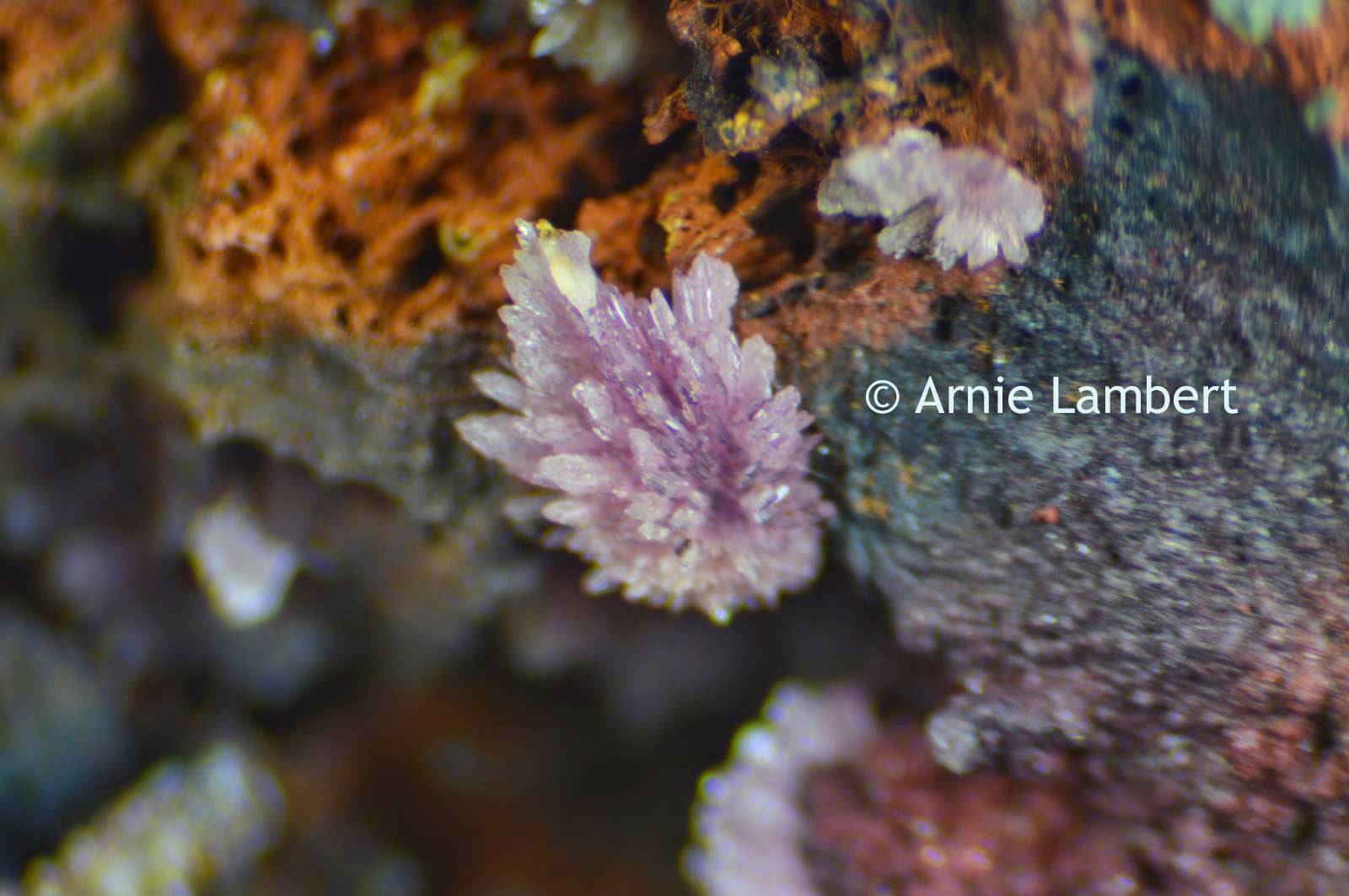
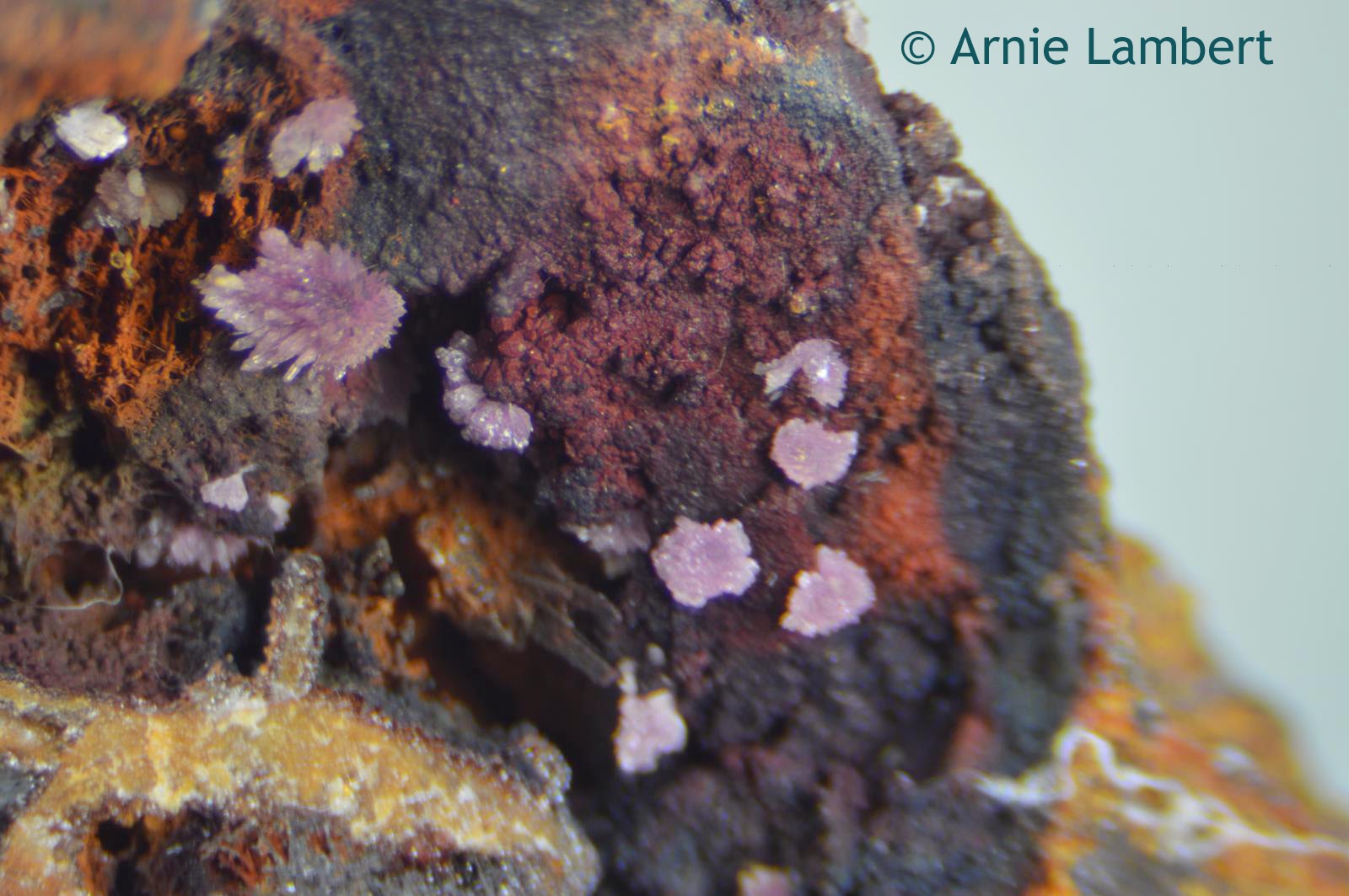
Minerals are best viewed under a stereo zoom microscope. A customer of Microscope World's, Mr. Arnie Lambert captured these mineral images using a stereo microscope and a Nikon SLR camera. The Nikon SLR camera was connected to the microscope through the eyetube on his microscope using the Nikon SLR camera microscope adapter.

Garnets in Rhyolite under the microscope, from New Mexico
Garnets in Rhyolite from New Mexico.
Garnets are a group of silicate minerals that have been used for many years as gemstones and abrasives. Garnet can be a variety of colors including red, orange, yellow, green, purple, brown, blue, pink, colorless and even black. The rarest color is blue. Rhyolite is an igneous, volcanic rock.

stengite under microscope from Indian Mountain
Strengite from Indian Mountain in Cherokee Co., Alabama
Stengite is a relatively rare iron phosphate mineral that is lavender, pink, colorless, or red. This mineral was named after Johann August Streng (1830-1897), a professor of Mineralogy at the University of Giessen, Germany.

Strengite from Alabama under the microscope
Strengite from Indian Mountain in Cherokee Co., Alabama.
Strengite minerals as seen under the microscope.
Strengite from Indian Mountain in Cherokee Co., Alabama.
Thank you Arnie for sharing your images with Microscope World!

Ranunculus under the Microscope

$
0
0
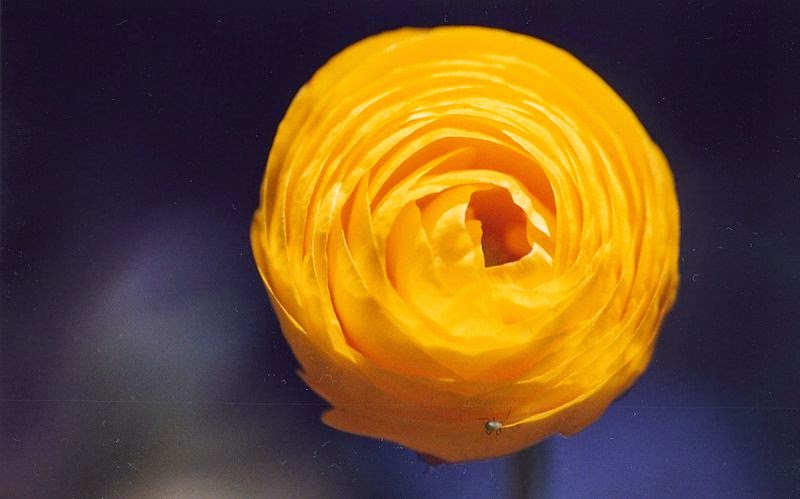
Ranunculus are a colorful genus of flowers that include buttercups, spearworts, water crowfoots and celandine. Typically Ranunculus have five petals, but occasionally more.
 
Yellow Flower
Ranunculus image courtesy of Joydeep Sen Sarma


The name Ranunculus is Latin for "little frog" and might refer to the many species of flowers found near water, similar to frogs.

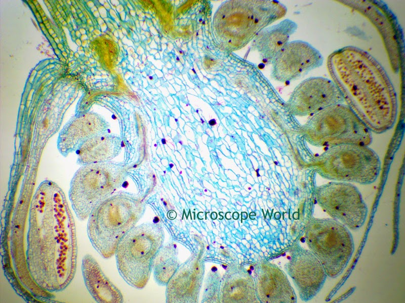
The images below of Ranunculus were captured under the U2 biological microscope using  a 5 megapixel microscope camera.

Ranunculus under the microscope at 40x
Ranunculus under the microscope at 40x magnification.

The Ranunculus prepared microscope slide is part of the Fruit and Flower microscope prepared slide kit available from Microscope World.

Ranculus under the microscope at 100x magnification.
Ranunculus under the microscope at 100x magnification.
Ranculus under the microscope at 400x magnification.
Ranunculus under the microscope at 400x magnification.

Capturing Microscope Images through an Eyetube

$
0
0
Ever wonder how it might be possible to capture images through your microscope if you don't have a trinocular port? There actually is a solution to this problem.

Replacing high school microscope eyepiece with c-mount camera adapter
The images below were all captured with a student microscope by removing the eyepiece, replacing it with the Generic C-Mount Adapter (AMA050) and threading the microscope camera onto the c-mount adapter.
Generic microscope c-mount adapter
Generic C-Mount Adapter (AMA050)
microscope camera
Microscope Camera
Microscopy image of laryxn.
Larynx captured under the microscope.

Larynx under the microscope.
The larynx microscope prepared slide is available for purchase as part of the Histology and Musculosketal Microscope Prepared Slide Kit.

Microscope Larynx Image
Larynx under the microscope.

Aphid captured under a biological microscope.
Aphid under the microscope.
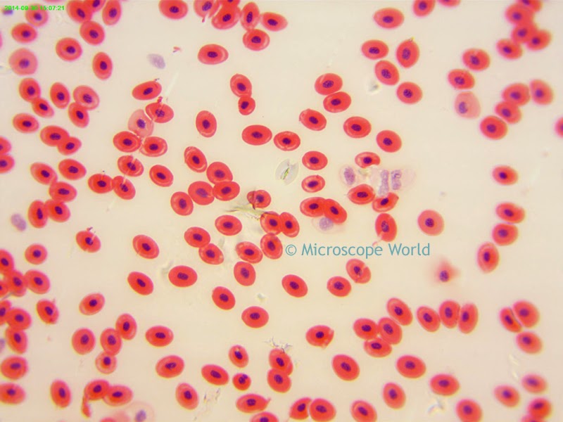
Student microscope image of frog's blood.
Frog's blood under the microscope.
If you have any questions about how to attach a camera to your microscope, send us an email!

Corn Seeds under the Microscope

$
0
0
Kernels of corn (or corn seeds) are used to make popcorn, feed animals, and make ethanol, among other uses. An average ear of corn contains 600 kernels on it.


The images below of a cross section of a corn seed were captured using a student biological microscope

Microscopy image of kernel of corn.
Corn seed captured under the microscope at 40x magnification.
The corn seed prepared microscope slide can be purchased as part of the Fruit and Flower Prepared Slide kit from Microscope World.

Microscope image of corn seed captured at 100x magnification.
Corn seed captured under the microscope at 100x magnification.
Corn seed captured under the microscope at 400x magnification.

Kids School Microscope Project Ideas

$
0
0
There are two types of microscopes you might find in your school, and each can be used to view different samples.

Student microscope
Student Biological Microscope F1

#1 - Biological Microscope

A biological microscope has light that shines up from beneath the stage directly through your sample and provides high magnification (40x, 100x, 400x). Samples are placed on a microscope slide and must be thin enough to allow light to pass through them. A cover slip is usually placed on top of the microscope slide.

Here are some great ideas of samples you can view with a biological student microscope.
  • Pond water (algae and bacteria, protozoans and other living organisms can be viewed).
  • Mold found on cheese such as Brie, Camembert or many other types of stinky cheese!
  • Hair - just a single strand of hair is all that is needed.
  • Cheek cells - use a Q-tip to scrape the inside of your mouth and place the sample on a slide.
  • Thinly sliced piece of an onion. Can you see any cells?
Stereo microscope for kids
Student stereo microscope with digital camera.

#2 Stereo Microscope

A student stereo microscope (or dissecting microscope) provides lower magnification (typically 10x - 40x) with light from above and/or below the sample. There is more room under a stereo microscope to place a larger sample such as a rock or plants.

Here are some great ideas of samples you can view in the classroom under a stereo microscope.
  • Dust, dirt and sand. The different grains and particles of sand and dirt are fun to view.
  • Textiles or fabric. What does the weave look like?
  • Printing text. What does a newspaper print look like compared to something you print from your computer?
  • Fruit and vegetables. Cut them thinly and then look at what you see under the microscope. Tomatoes, mushrooms and kiwi are all interesting to look at.
  • Coins and dollar bills.
  • Flowers and insects.
Insects under microscope.
Insect captured at 45x magnification.
Have fun with your microscope. If you capture any interesting images send us an email, we would love to see them!

Spleen Under the Microscope

$
0
0
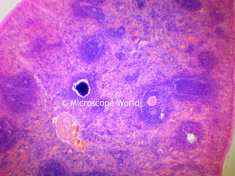
Just about all vertebrates have an organ called the "spleen". The spleen has a similar structure to a large lymph node and acts primarily as a blood filter. The spleen plays important roles in regard to red blood cells and the immune system. The spleen removes old blood cells and holds a reserve of blood, which can be valuable in the case of shock. The spleen also synthesizes antibodies in its white pulp and removes antibody-coated bacteria and antibody-coated blood cells by way of blood and lymph node circulation.

Image courtesy US Government
The images below were captured under the U2 biological microscope using a microscope digital camera.

Spleen under microscope at 40x magnification.
Spleen captured under the microscope at 40x magnification.
Spleen under the microscope at 100x magnification.
Spleen captured under the microscope at 100x magnification.
Spleen captured under microscope at 400x.
Spleen captured under the microscope at 400x magnification.

Tuberculosis Viewed Under the Microscope

$
0
0
Tuberculosis (TB) is an infectious disease caused by various strains of mycobacteria. Tuberculosis usually attacks the lungs and it is spread through the air when people with an active TB infection cough or sneeze. Symptoms of TB include chronic cough, often with blood-tinged sputum, fever, night sweats and weight loss. Diagnosis of TB relies on radiology (chest X-Rays), as well as microscope examination. One -third of the world's population are thought to have been infected with TB with new infections occurring in about one percent of the population each year.

The microscope prepared slide of Tuberculosis used to capture the images below can be found in the Bacteriology Prepared Slide Kit.

TB under the microscope at 40x
Tuberculosis under the microscope, 40x magnification.
These images of TB were captured using the U2 digital microscope (5mp camera).

TB under the microscope at 100x magnification.
Tuberculosis under the microscope, 100x magnification.

TB under the microscope, 400x
Tuberculosis under the microscope, 400x magnification.

HD Digital Stereo Microscope Images

$
0
0
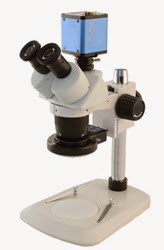
Stereo Microscope 10x 30x with HD camera
S2-TPHD Stereo Microscope
The Richter Optica S2-TPHD is a 10x / 30x stereo microscope with a high definition camera. Right now this microscope system is on closeout special and has been reduced by $200 to $799 with free shipping. This past week Microscope World captured some images with this specific setup. The LED ring light provides plenty of light and the camera captured some good images. The camera allows a live view on an LCD projector or monitor using the HDMI cable, or capture of 5 megapixel images to an SD card.


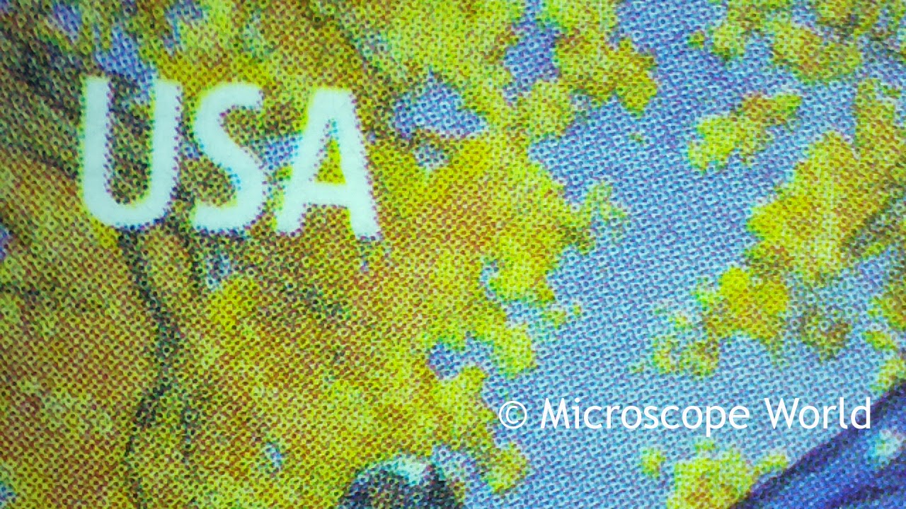
Stamp image under microscope
Postage stamp captured under the microscope at 10x magnification.

Steel under microscope at 10x
Steel Blade captured under microscope at 10x.

Printed Circuit board under microscope at 10x
Printed Circuit Board under microscope at 10x.


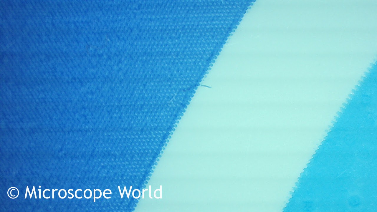
Plastic film at 10x under stereo microscope.
Plastic Film captured under microscope at 10x.

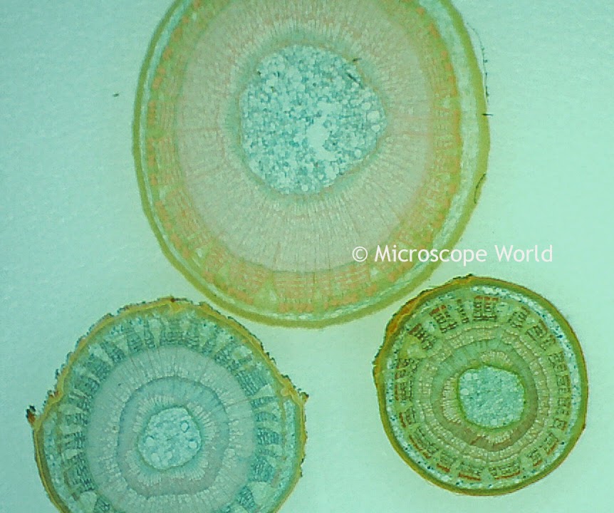
Monocot and dicot under microscope at 10x
Monocot & Dicot captured under microscope at 10x.

Postage stamp under microscope at 10x magnification.
Postage stamp under microscope at 10x.

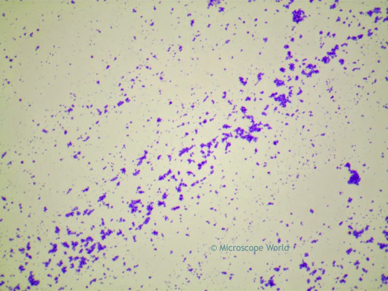
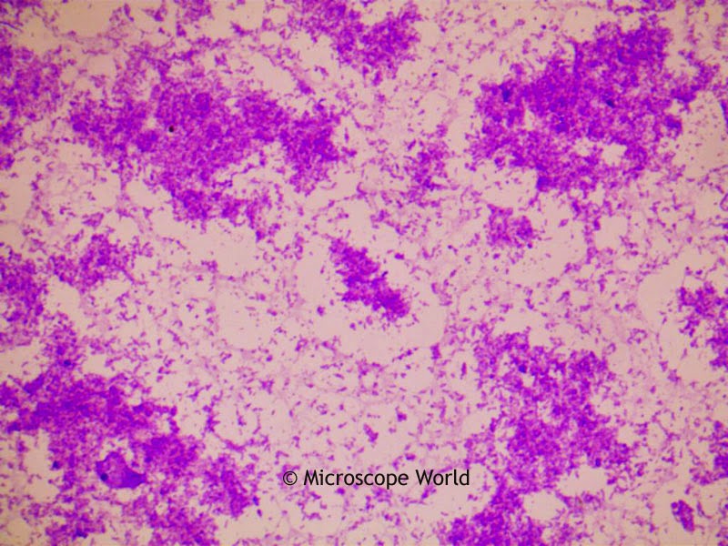
Bacteria under the Microscope

Pollen under the Microscope

$
0
0
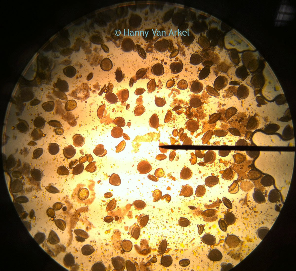
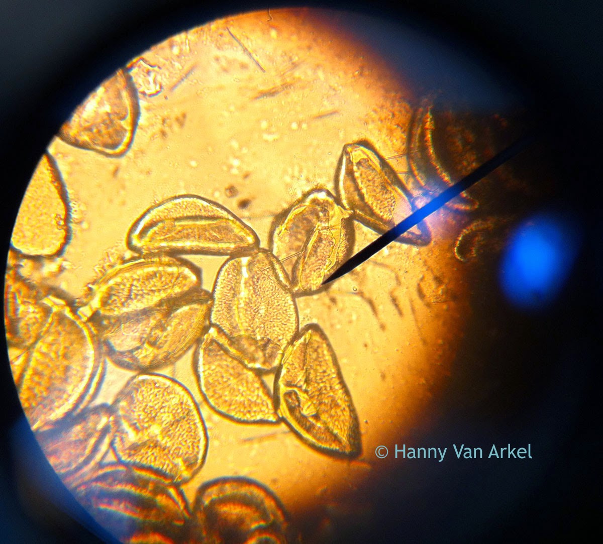
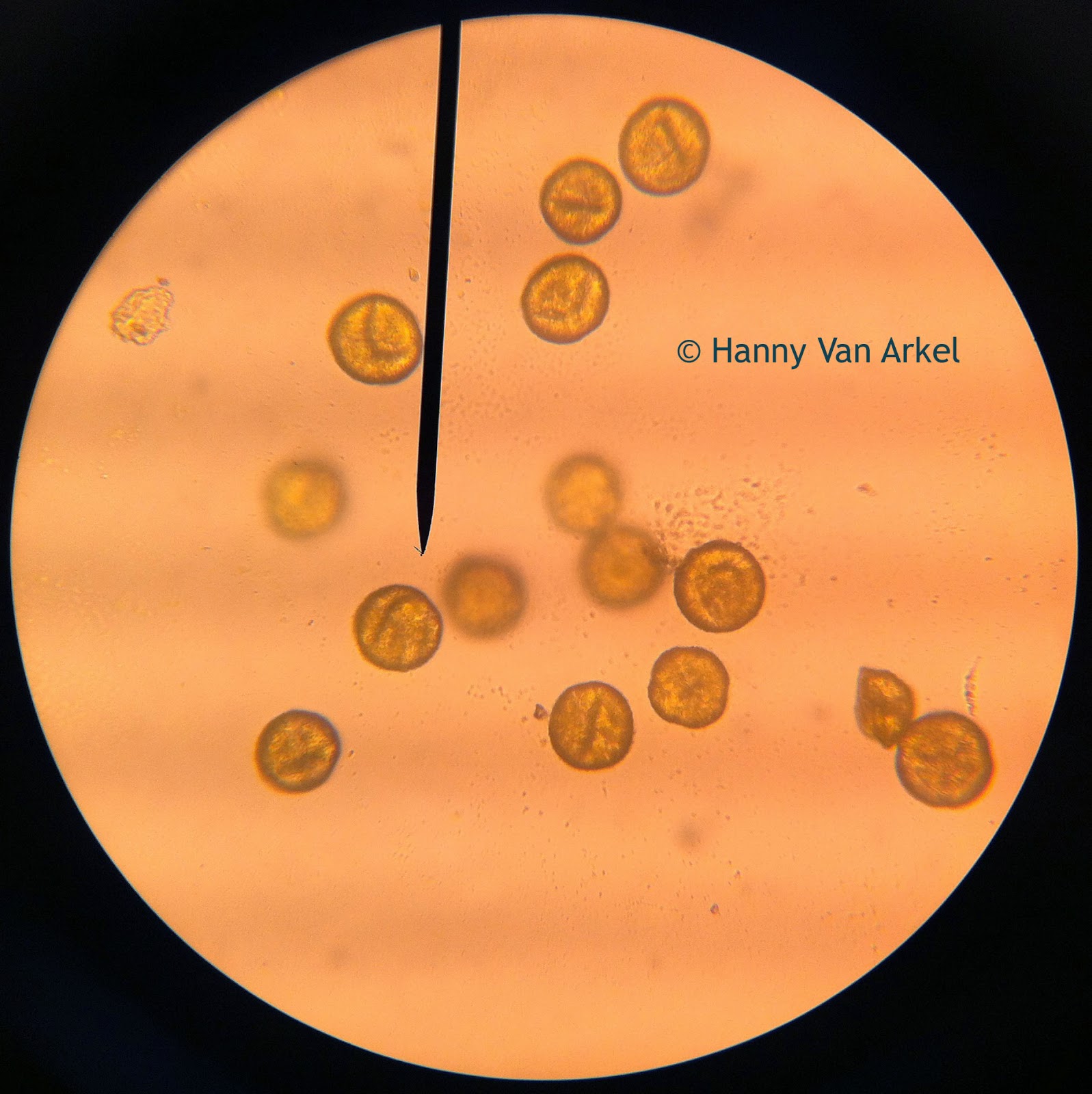
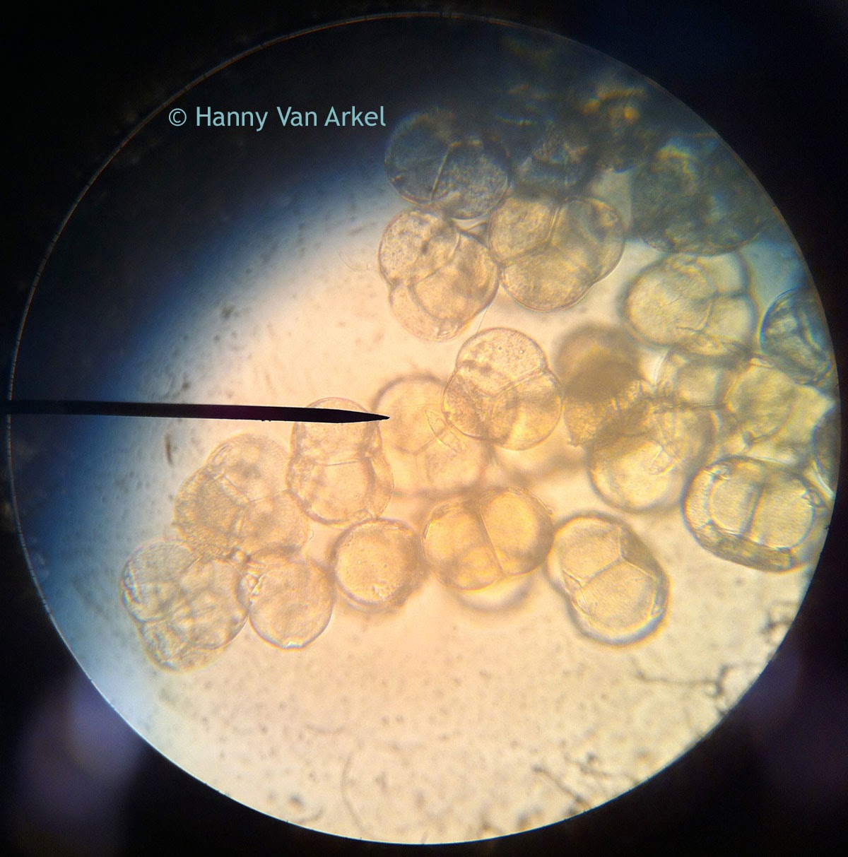
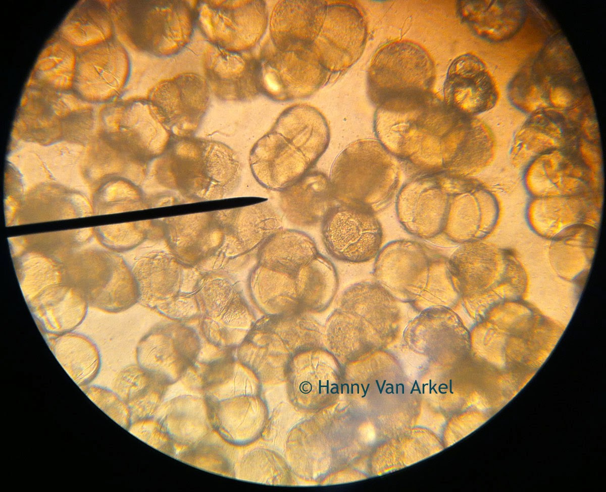
The following images of pollen were captured by Hanny van Arkel, a biology teacher in the Netherlands, using a student microscope. You can view more of her pollen images here! The flowers with the pollen on them were brought into class by Hanny's twenty-six 13-year-old students.

Pollen captured under microscope at 100x
Pollen under the microscope at 100x magnification.
Pollen microscopy image at 400x
Pollen under the microscope at 400x.
pollen microscopy image at 400x
Pollen under the microscope at 400x.
Pollen under the microscope at 400x
Pollen under the microscope at 400x.
Pollen at 400x under microscope
Pollen under the microscope at 400x.
Thank you to Hanny van Arkel and her entire Biology Class for sharing these microscopy pollen images. If you have some images you would like to share, please email us.
Viewing all 422 articles
Browse latest View live