Quantcast
Channel: Microscope World Blog
Viewing all 422 articles
Browse latest View live

Phytoplankton under the Microscope

$
0
0
The ocean is vast and today we are going to focus specifically on Phytoplankton under the microscope. The term Phytoplankton comes from the Greek word "Planktos" which means wanderer or drifter, exactly what Phytoplankton do in the ocean, as they are unable to swim against major ocean currents.

Phytoplankton thrive near the surface of the ocean since they are organisms that make food from sunlight (known as photosynthesis). Through the process of photosynthesis, Phytoplankton play a large role in the ocean, which contributes to climate and the air breathed each day! Phytoplankton produce about fifty percent of the oxygen you breathe every day, and Diatoms (a single-celled type of Phytoplankton) also help reduce greenhouse gasses. Scientists collect Phytoplankton from the ocean using nets in order to study the organisms.

There are a number of ocean animals' life cycle that depends on Phytoplankton for their existence.

Ocean life cycle including photoplankton.
Example of the ocean life cycle involving Phytoplankton.
In one single teaspoon of ocean water you might find over a million Phytoplankton. When conditions are ideal, Phytoplankton can grow in such massive numbers that the bloom can be seen from outer space.

Diatom under the biological microscope.
Diatom under a biological microscope.
Diatoms are another type of plant-like plankton that come in varying shapes including zigzags, ribbons, and fans. They have a protective cell wall made of glass, and their spines help prevent them from sinking. Diatoms also form chains which help keep them near the surface.

You will need the following items to view plankton with the microscope:
  • Plankton - visit a local pond, stream or the ocean. Gather some water, preferably with larger particles in it, look for pieces of seaweed, kelp or plants.
  • Petri Dish
  • Toothpicks to move the plankton around while viewing.
  • Stereo Microscope
  • Optional: Microscope camera for capturing images.
Place your sample water in a jar and leave it in a sunny area. Pour some water into your petri dish. The more water in the petri dish means that any plankton will move around more and you may be able to observe them swimming. Less water in the dish makes for easier viewing, as the plankton will not move around as much and therefore won't swim out of your field of view.

When viewing the plankton under the stereo microscope you may need to switch between incident and transmitted light in order to get the best view. Using a frosted glass stage plate is usually most effective for observing transparent specimens such as plankton. If you are primarily using light from above, switch to the black or white stage plate.

Finally, if you have access to a biological microscope, you may want to put a small sample of water on a depression slide with a cover slip and look for Phytoplankton at a higher magnification.

Phytoplankton under the microscope at 40x.
Phytoplankton captured under the microscope at 40x magnification.
Source: Center for Microbial Oceanography: Research and Education (C-MORE) is a Science and Technology Center headquartered at the University of Hawaii. Parts of this post are from the Microscope in Middle Schools Project. They have an exceptional collection of Phytoplankton images.

Insect under the Microscope

Top 10 Holiday Science Geek Gift Ideas

$
0
0
Have someone in your life who loves science? Here are top ten great science gift ideas for the science geek, both young and old.

Glass Galileo Thermometer

The Galileo thermometer consists of a sealed glass tube that is filled with water and several floating bubbles. The bubbles are glass spheres filled with a colored liquid mixture. This liquid mixture may contain alcohol, or it might simply be water with food coloring. Attached to each bubble is a little metal tag that indicates a temperature. A number and degree symbol are engraved on the tag. Learn more about the glass Galileo thermometer here.

Glow in the Dark Constellations

Select from a number of different constellations and transform your ceiling into an astronomy learning room! These constellations are especially fun for kids, as they can learn a bit about astronomy when they go to bed each night. View a number of constellation options here.


Vacuum Coffee Brewer

No other coffee maker has fascinated passionate coffee drinkers like this one. Its exceptionally effective vacuum brewing method extracts all precious oils of your favorite coffee. The vacuum brewing process is entirely sealed off so no aroma can escape. In addition, the brewing time and temperature are perfectly calibrated, ensuring a perfect cup of coffee with each brew. Plus, it just looks scientific!

Kids microscope gift ideasMicroscope

Whether you are young at heart, or looking for a gift for a kid, microscopes make fun holiday gifts for the science geek in your life. Biological microscopes are perfect for viewing slides, bacteria in pond water, or the nuclei in your cheek cells. A stereo dissecting microscope is great for viewing flowers, insects, or even dissecting a frog. View a microscope gift guide here.


 Test Tube Spice Rack

Dean and Deluca makes a very nice (and slightly pricey) test tube spice rack that is perfect for any scientist who also loves to cook. We also found some great instructions on how to make your own test tube spice rack here. If you have a bit of time, you can make your own test tube spice rack and customize the labels with your own personal touch.

Science Museum Gift Cards

Most local science museums offer gift cards for either entry into the museum, or to the museum gift shop. Many times outings are more memorable than tangible gifts, and this could be a gift for the entire family.

Da Vinci's Catapult Wood Kit

No matter what age you are shopping for, this gift is a winner. Da Vinci's Catapult Wood Kit takes over an hour to assemble and will catapult small items up to 100 feet. This honestly might be a great gift for the executive in your life who spends some frustrating hours at a desk. Probably not a good gift for small children, as there are too many small parts that could be a choking hazard. You can find Da Vinci's kit here.

Flower prepared slide is part of the microscope prepared slide kit from Microscope World.Microscope Prepared Slide Kit

Know someone who already has a microscope? An inexpensive and fun gift is simply a new prepared slide kit. With eight kits to choose from covering everything from botany, zoology, histology and bacteriology, you're sure to find a microscope prepared slide kit for the scientist in your life.


Low Temp Stirling Engine Model

This little stainless engine can be used to teach teenagers and older students how heat engines work. Or for the scientist with a desk, the engine is fun to watch. This low-temperature-differential Stirling engine works with hot water, ice water, a combination of hot water with ice cubes, or a hair dryer. The bigger the temperature differential the faster it runs. You can view Stirling engines here.

Super Strong Rare Earth Magnets

Rare-earth magnets are strong permanent magnets made from alloys of rare earth elements. Developed in the 1970s and '80s, rare-earth magnets are the strongest type of permanent magnets made, producing significantly stronger magnetic fields than other types such as ferrite or alnico magnets. The term "rare earth" can be misleading as these metals are not particularly rare or precious; they are about as abundant as tin or lead. You can find rare earth magnets here.

Happy holiday scientist shopping!


Motic Trace Forensic Microscope Software

$
0
0
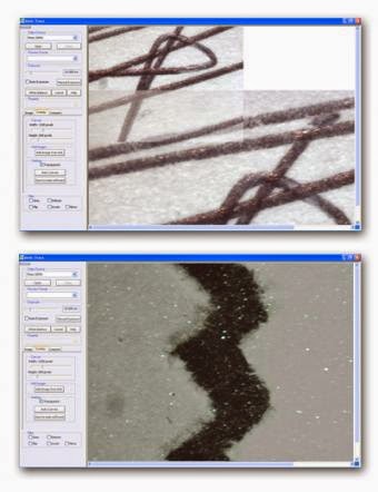
Motic Trace is a forensic software that removes the need to purchase expensive comparison microscopes. Simply connect two or more Motic microscope cameras or Motic digital microscopes to your computer and get immediate live comparison imaging. Turn your existing microscopes into comparison microscopes. This is a perfect solution for forensic science teaching or forensic departments with a limited budget.

Motic Trace forensic microscope comparison software.
Motic Trace Software

  • Compare images side by side.
  • Overlay several live images.
  • Resize & rotate images independently.
  • Match fingerprints from live or saved images.
  • Check handwriting for authenticity.
  • Compare two pieces of fabric.
Simply connect two or more Moticam cameras or Motic digital microscopes to the computer and get an immediate live comparison through the Motic Trace Software. The software allows you to create reports with accurate measurements and editing tools.

Marijuana Microscope

$
0
0
Growing marijuana or identifying marijuana is often made easier with the use of a microscope. The marijuana microscopes shown below are popular among marijuana growers and marijuana inspectors.

Cannabis leaf, Cannabis Sativa
Photo: Jon Richfield

Handheld USB Marijuana Microscope

The handheld USB microscope is a great tool for viewing buds while they are still on the marijuana plant. This is helpful for determining when to harvest the plant. The handheld USB microscope shows a live image on the computer and you can capture and save the images.


Stereo zoom marijuana microscopeStereo Marijuana Microscope


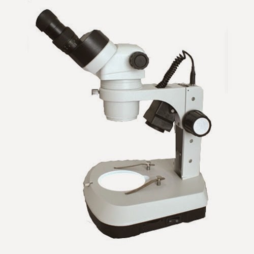
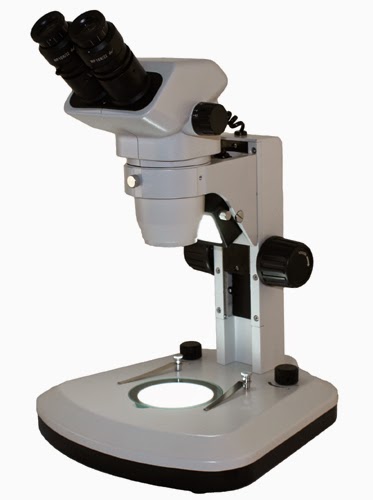
The stereo microscope is available with either dual magnification (less expensive and 10x and 30x is usually the best combination of magnification) or zoom magnification (around 10x - 40x).Using a stereo microscope is one of the best ways to view buds and leaves. The LED light is a good illuminator to use as it does not heat up and won't damage the plants.


Digital LCD stereo marijuana microscope
Digital Stereo Marijuana Microscope

Digital stereo microscopes are available with either a tablet LCD display (simple to use and easy to capture and save images), or a microscope USB camera connected to the marijuana microscope. Digital microscopes allow you to capture and easily email images, or even make measurements with the software, add annotations to images and save them.

Lactobacillus under the Microscope

$
0
0
Lactobacillus is a genus of gram-positive rod-shaped bacteria that is a major part of the lactic acid bacteria group. Most bacteria in this group convert lactose and other sugars to lactic acid. In humans Lactobacillus is present in the vagina and the GI tract, where they make up a small portion of the gut flora. Typically Lactobacillus bacteria are benign, except in the mouth where they have been known to cause cavities and tooth decay.

Lactobacillus bacteria under the microscope at 40x magnification.
Lactobacillus under the microscope at 40x magnification.
These images of Lactobacillus were captured with a Richter Optica high power compound microscope using a microscope digital camera.

Bacteria captured under a Richter Optica microscope at 100x magnification.
Lactobacillus captured under the microscope at 100x magnification.
The Lactobacillus rod-shaped bacteria is part of the bacteriology microscope prepared slide kit.

Bacteria under the microscope at 400x magnification.
Lactobacillus under the microscope at 400x magnification.

Cyber Monday Microscope Special

Mud Logging Microscopes

$
0
0
Mud logging is the creation of a detailed record (known as a well log) of a borehole by examining the cuttings of rock brought to the surface of the earth with a circulating drill. Mud logging is commonly used in petroleum exploration, but it is also sometimes used when drilling wells for water or other mineral exploration.

Mud logging tracks the following:
  • Rate of penetration - the rate at which the drill bit breaks the rock under it when it deepens the borehole.
  • Porosity - the pore space within the rock structure. A good analogy is to think of the holes in a sponge, oil and gas resides within this space.
  • Lithology - color, texture, grain size of the sample.
  • Gas - heavy gases such as ethane, propane, and butane are detected to help determine the type of oil or gas that is contained in the location.

Mud logging microscopes are used to measure the lithology (color, texture, grain size) of the sample, usually at a lower magnification (10x - 20x). Below is a sample of drill cuttings of shale from drilling an oil well in Louisiana. In the image the sand grain and red shale are about 2mm in diameter.

Credit: Mud Engineer / Wikipedia
The sample above would be considered about 90% shale, 5% sand and 5% lime when listed in the mud log.

well logging microscope
Mud Logging Microscope
Mud logging microscopes provide magnification in the range of 10x - 45x and typically utilize either a ring light or a built in LED light on the stand. Some mud logging companies prefer a microscope with a camera in order to easily document their findings. Pseudo-darkfield illumination is sometimes used to bring out features of a specimen not normally seen under brightfield light. You can view mud logging microscopes here.

Snowflakes under the Microscope

$
0
0
When snowflakes fall on mittens or coats, it is fascinating to view the unique intricate details and designs of each one. Wilson A. Bentley lived in Jericho, Vermont (1865-1931) and was much ahead of his time in discovering that "no two snowflakes are alike."

Bentley was a self-educated farmer and attracted attention from around the world with his pioneering photomicrography of snow crystals. He adapted a microscope to a bellows camera and became the first person to photograph a single snow crystal in 1885.

Wilson Bentley

Throughout his lifetime Bentley captured over 5000 snowflakes, never finding two alike. Over the years his photographs were acquired by Universities throughout the world and have been published in many articles and magazines.

Wilson Bentley snowflake, image courtesy Snowflake Bentley
Wilson Bentley wrote an article for Popular Mechanics Magazine in 1922 about photographing snowflakes. Some of the hardest part of his work involved being in the cold temperatures (blizzards) that produce beautiful snowflakes. You can read Wilson Bentley's "Photographing Snowflakes" article here.

Wilson Bentley snowflake, image courtesy Snowflake Bentley
Wilson Bentley used a compound microscope (high power biological microscope) to view and photograph the snowflakes. However, there were no digital microscopes back when Bentley performed his snowflake photomicrography. He was creative and attached his bellows camera to the microscope in order to capture the beautiful images.

iPad Biological Microscope

$
0
0
Microscope World is excited to introduce the iPad mini Biological laboratory microscope package.

Biology iPad digital microscope and WiFi camera system.
U2iPad Biological Digital Microscope
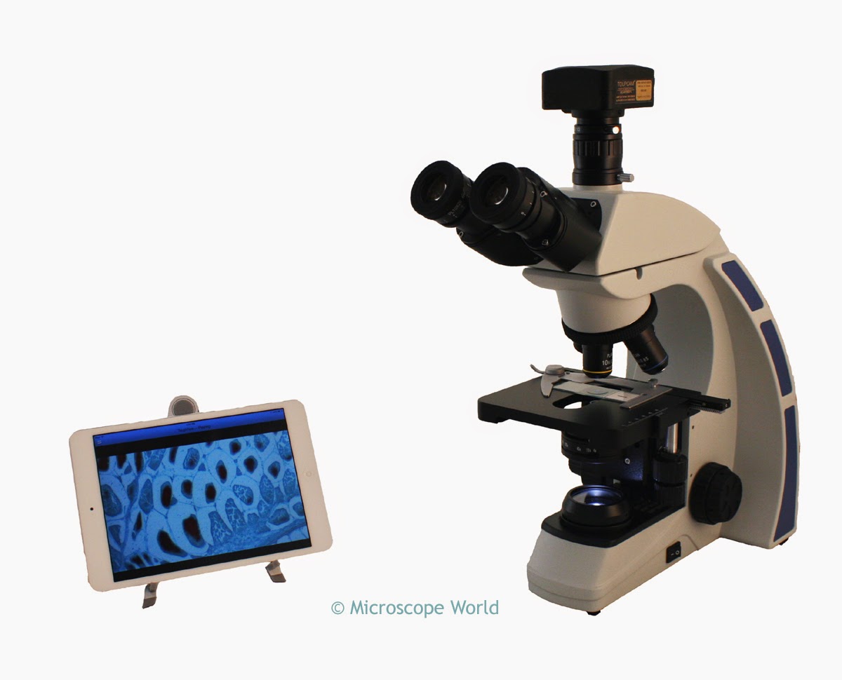
The digital biological microscope system includes an iPad Mini 2 tablet with the microscopy App pre-installed, WiFi camera with its own network, and a laboratory biological microscope that provides 40x, 100x, 400x and 1000x magnification. 

View live images from the microscope directly on the iPad. The tools included in the Software App allow for adjustment of color, exposure and gain, and white balance. Images can be captured and saved directly onto the iPad. When not using the microscope, the iPad can be used around the lab, office or home for other uses.

Beautiful Wheat under the Microscope

Industrial iPad Microscope

$
0
0
Microscope World is excited to introduce our newest Industrial iPad Microscope system. This stereo zoom microscope has 7x - 46x continuous zoom magnification and is outfitted with a WiFi camera that provides a live image directly to the iPad. Images can be captured and saved on the iPad, and adjustments can be made to color, exposure, and gain.

iPad Stereo Zoom Microscope System with Flexible Clamping Arm

The iPad stereo zoom system is available with two different iPad stand options. A stand that clamps to the table and is flexible for positioning the iPad right at eye level near the microscope is available.

The alternate stand available is a small metal table stand that can hold the iPad near the microscope, or a bit further away from the working area.


iPad Stereo Zoom Microscope System

If you already have an iPad and would like to use a microscope system with your iPad email us or call us at 800-942-0528 and we would be happy to help you!

Pear under the Microscope

$
0
0
There is evidence of the pear being used as a food since prehistoric times. Many traces of it have been found in Swiss lake-dwellings and the word "pear" occurs in Celtic languages. There is even a recipe for a spiced, stewed-pear patina (or souffle) in the Roman cookbook attributed to Apicius.

Photo: USDA Keith Weller
The images below are of a cross section of a pear and were captured using a high school biological microscope.  The cross section of pear prepared slide is part of the Fruit and Flower Slide Kit.

Pear prepared slide under the microscope at 40x.
Cross section of a pear captured under the microscope at 40x magnification.
Pear under the microscope at 100x magnification.
Cross section of a pear captured under the microscope at 100x magnification.
Pear under a biological microscope at 400x magnification.
Cross section of a pear captured under the microscope at 400x magnification.

Sugar Swizzle Stick Science Project

$
0
0
Have you ever looked at sugar under the microscope? Under a stereo microscope, sugar looks like this:

Sugar under a stereo microscope at 40x.
Sugar crystals under a stereo microscope at 40x magnification.
Sugar captured under a polarizing microscope looks like this:

Sugar under a polarizing microscope at 200x.
Sugar under a polarizing microscope at 200x magnification.
A fun student science project will allow you to make your own sugar sticks (and look at sugar crystals under the microscope!)

Photo: Deisy Mejía
You will need these supplies:
  • Wooden skewer
  • Clothes pin
  • Tall glass
  • 3 cups of sugar
  • 1 cup of water
Measure the wooden skewer so that when you hold it straight down into the glass it does not quite touch the bottom of the glass. You will use the clothespin to secure it across the top of the glass. Set the clothespin and skewer aside.

Find an adult to help you boil your water and add about 1/4 cup of the sugar, stirring as it dissolves. Each time the sugar dissolves, slowly add more. Continue stirring. Each time you add more sugar it will take longer and longer for it to dissolve. Keep stirring! Once no more sugar will dissolve, turn the heat off and let it cool for about 20 minutes.

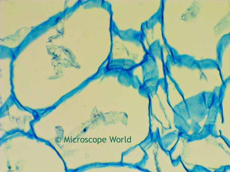
Have the adult pour the hot liquid into the tall glass. Now insert the skewer into the middle of the glass and secure it at the top with the clothespin, so it hangs straight down. Over the next week sugar crystals will form on your skewer!

If you want to make colored sugar swizzle sticks, add a few drops of food coloring to your mixture. Once you are finished making your swizzle stick, break off a small piece of rock sugar and look at it under the microscope. What do your crystals look like? If you capture some good photos email us and we will share them on our Facebook page!

Lily Flowers under the Microscope

$
0
0
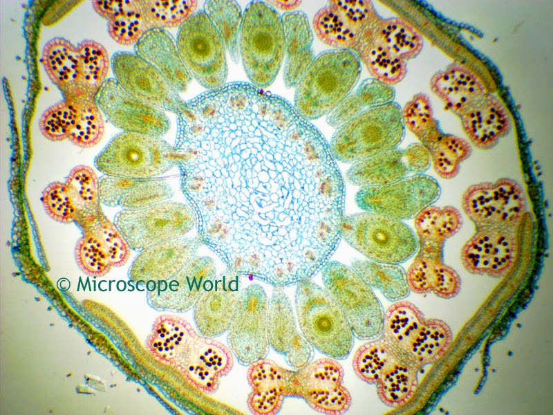
Lilium is a genus of herbaceous flowering plants grown from bulbs, all with large prominent flowers.

Photo: Stan Shebs
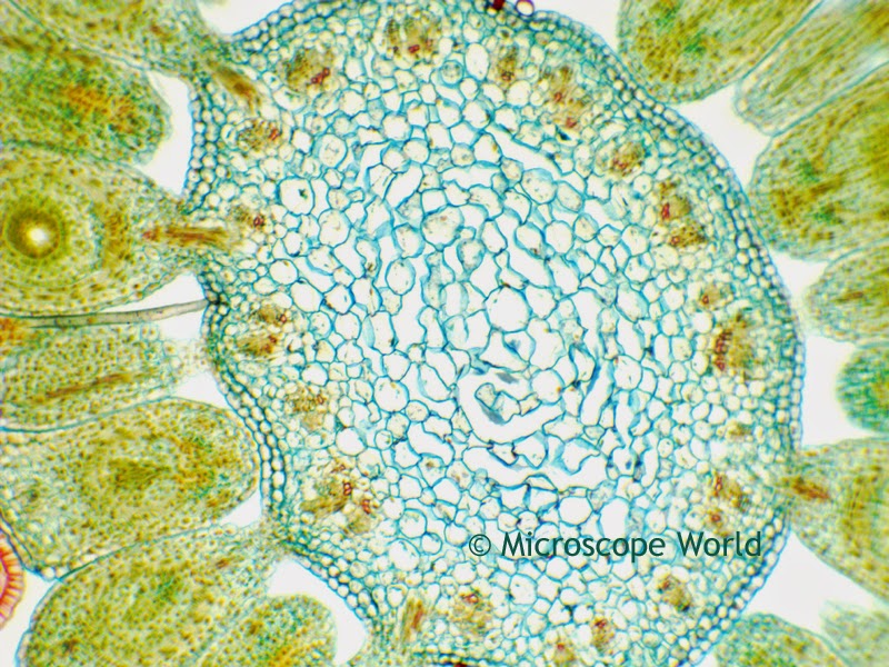
The following images are of a cross sections of Lilium, part of the Fruit & Flower Prepared Slide Kit. The microscopy images were captured using a Richter Optica U2 biological microscope along with the DCM3.1 microscope camera (3.2 megapixels).
Lily captured under the microscope at 40x magnification.
Lilium captured at 40x under the microscope.

Notice in the image above the center looks to be shaped like a Lily!
Lily captured at 100x under the microscope.
Lilium captured at 100x under the microscope.
Lilium captured at 400x under the microscope.


What is a Compound Microscope?

$
0
0
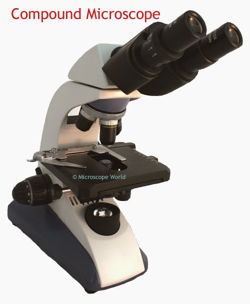
A compound microscope is another name for a high power or biological microscope. A compound microscope will typically have magnifications of 40x, 100x, 400x and sometimes 1000x. A microscope advertising magnification above 1000x is simply providing empty magnification, and images above 1000x will not be clear. Blood cells, protozoans and bacteria can all be viewed at 400x magnification and it is a common misconception that more magnification is better.

Compound microscope high power objectives.
Compound Microscope High Power Objective Lenses

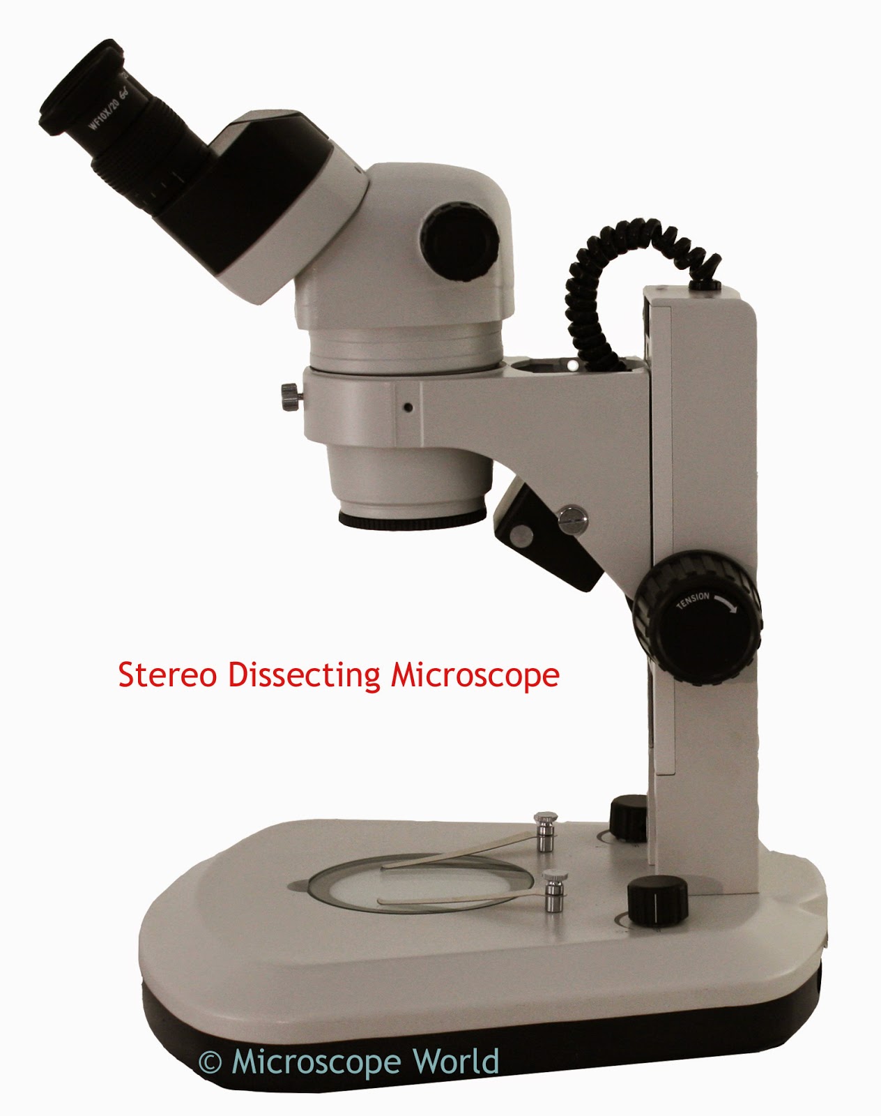
Students typically use either a compound microscope in biology class, or a stereo dissecting microscope. A stereo dissecting microscope provides lower magnification (typically 10x - 40x) and is used to view anything you might hold in the palm of your hand such as coins, flowers, insects, or a frog. A compound microscope requires the use of slides and cover slips for viewing samples the naked eye can not see such as bacteria or cells.

Stereo dissecting microscope 10x-40x
Stereo Dissecting Microscope

Compound microscope with high magnification
Compound Microscope

Melon under the Microscope

$
0
0
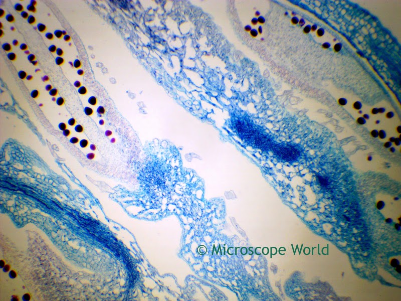
Cucumis is a genus of twining, tendril-bearing plants in the Cucurbitaceae family which includes the cucumber, cantaloupe and honeydew melon as well as the sugar and winter melon. The images below of Cucumis Staminate Flowers were captured using the Richter Optica U2 biological microscope along with a 3 megapixel microscope camera. The microscope prepared slide is part of the Fruit & Flower Prepared Slide Kit.

Cucumis staminate flower under the microscope at 40x.
Cucumis Staminate Flower under the microscope at 40x magnification.

Cucumis Staminate flower under the microscope at 100x.
Cucumis Staminate Flower under the microscope at 100x magnification.

Flower under a biological microscope at 400x magnification.
Cucumis Staminate Flower under the microscope at 400x magnification.

What is a Microscope Eyepiece Reticle?

$
0
0
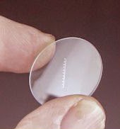
Microscope eyepiece reticles are small circular disks that have a ruler, grid or some other measuring format printed on them that can be inserted into a microscope eyepiece. When you look through the eyepiece once the reticle is installed, the ruler or grid is imposed upon your microscopy image.

How to determine if your eyepiece accepts a reticle:

microscope eyepiece
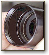
Bottom of the eyepice.

Not all microscope eyepieces are fitted to accept a reticle. In order to figure out if yours does, follow these instructions.

Remove your eyepiece from the microscope and look into the bottom of it. If your eyepiece accepts a measuring reticle, you will see a retaining ring that can be removed.


How to measure the correct diameter of reticle required:


microcope reticle retaining ring
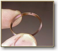
Reticle Retaining Ring
If you see a retaining ring, go ahead and remove the ring. You will then want to measure (in mm) the diameter of the glass reticle that the eyepiece will hold. This is an important measurement as many eyepiece reticles are custom printed and can not be returned. Keep in mind that if you measure the retaining ring diameter, this may be 1mm larger than the size reticle requierd, as the glass reticle must be able to drop onto the shelf in the eyepiece, while the retaining ring will simply hold it in place. Microscope eyepiece reticle sizes are anywhere from 16mm - 28mm in diameter.


How to determine what type of microscope reticle to purchase:

Microscope Eyepiece Reticle
Microscope Reticle
Once you know that your microscope eyepiece can accept a reticle, now it is time to determine what type will best suit your needs. The most common types of reticles are ruler reticles, crossline reticles and grid reticles. For basic measurement a ruler reticle will work well.

Determine if you would like to measure in mm or inches. Also, keep in mind that when using a ruler reticle, the distance that is between the lines when looking through the microscope follows this formula:

Reticle Division ÷  Objective Lens Value = Distance Between Lines

The magnification of the eyepiece itself has no affect on the reticle divisions you will view. For example, if your ruler reticle is 10mm with 100 divisions, your reticle division is 10 ÷ 100 = 0.1mm.

When using the 4x objective to make measurements with the above mentioned reticle, your distance between lines when measuring is: 0.1mm ÷ 4 = 0.025mm.

When using that same reticle with the 100x objective lens the distance between lines is 0.1mm ÷ 100 = 0.001mm.

Many specialized microscope reticles are available including comparator reticles, grain sizing reticles and concentric circles.

Once you have purchased your reticle, you will want to calibrate your microscope. Learn more about calibrating your microscope here.

Beautiful Microscopy Images

Best Student Microscopes

$
0
0
There are several important factors to look into when purchasing a student microscope. Quality is important and you can find a great article here about evaluating microscope quality. Other than a high quality microscope, here are a few factors that are important:
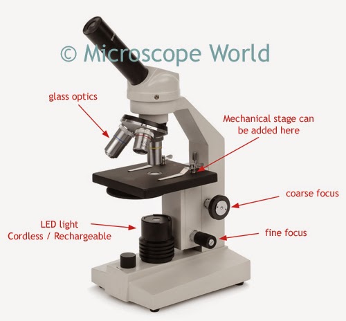
  • Both coarse AND fine focusing. Once you are viewing specimens at 100x magnification or higher, having a fine focusing knob is essential for getting a crisp and clearly focused image. Inexpensive kids microscopes will only have a coarse focusing knob. The fine focusing knob eliminates quite a lot of frustration for both children and adults.
  • Consider the light. Both LED and halogen light sources are cool. If you are looking at living samples, this is the best way to keep those samples alive! LED bulbs last much longer than any other type of bulb. Fluorescent and tungsten lights heat up when in use. Additionally, is it important for you to have a cordless microscope? Many microscopes now are rechargeable and this makes them more kid-friendly, as you don't have cords stretched from a table to a wall outlet when the microscope is in use.
  • Do you need to capture images? There are a number of digital kids microscopes available from WiFi microscopes, and LCD tablet microscopes, to USB digital microscopes. If you want to capture images, decide whether you would prefer to view your images on a computer next to your microscope, or on a tablet mounted directly on your microscope. Keep in mind that you can often convert a standard microscope to digital after purchasing, if you don't wish to invest in a digital microscope up front.
  • Consider the Optics. To be completely honest - any kind of plastic optics are horrible. Eyepieces and objective lenses should all be made of glass. If you are looking at a kids microscope under $50 it probably has plastic optics (as well as plastic parts, which break easily). Looking through plastic optics is similar to looking through an extremely dirty window. 
  • Consider the Magnification. Anything above 1000x in empty magnification and is unnecessary. You can view bacteria, blood cells and Protozoans all at 400x. More magnification is not better.

Best Student Microscopes

Richter Optica F1 kids microscope
F1 Student Microscope
#1 - Best Kids Microscope:

F1 Student Microscope


#2 - Best High School Microscopes:
Richter Optica HS-1M high school student microscope features.
HS-1M High School Microscope

HS-1M Basic High School Microscope


UX-1 Plan Achromat High School Microscope

Richter Optica UX-1 Plan Achromat High School Microscope
UX-1 Plan Achromat Microscope


#3 - Best Digital Microscopes:

UX1-LCD Tablet Microscope for High School
UX1-LCD Tablet Microscope
UX1-LCD Digital Tablet LCD Microscope

  • Coarse & Fine Focusing
  • Cordless, Rechargeable, LED Light
  • Glass Optics, Metal Parts
  • Includes Mechanical Stage
  • Includes Touch-Screen Android Tablet
  • 40x, 100x, 400x, 1000x
  • Plan Achromat Objective Lenses
  • Microscope Operates with or without use of Tablet
  • Capture and save images and video
If you are looking for a specific student microscope setup please contact us for further information.
Viewing all 422 articles
Browse latest View live