Quantcast
Channel: Microscope World Blog
Viewing all 422 articles
Browse latest View live

Plant Seed Embryo Under the Microscope

$
0
0
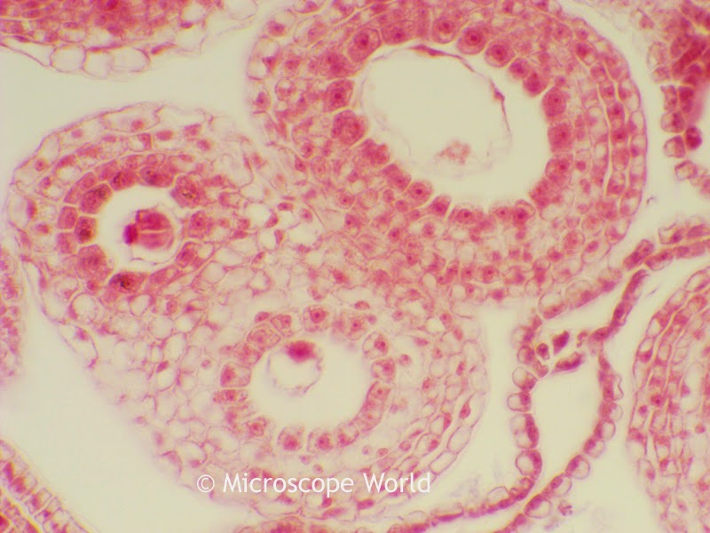
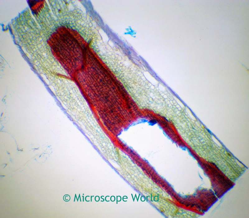
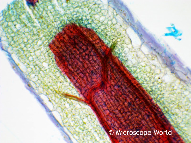
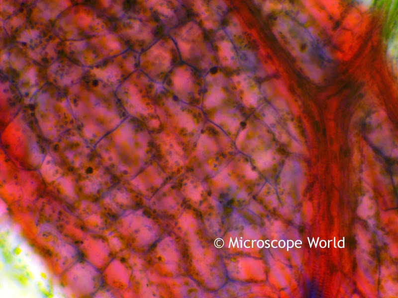
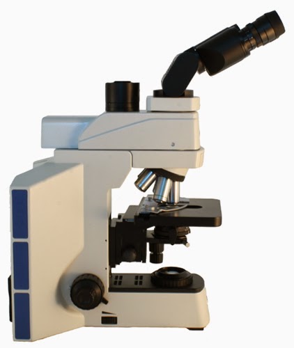
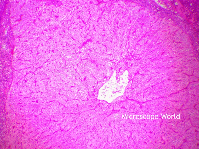
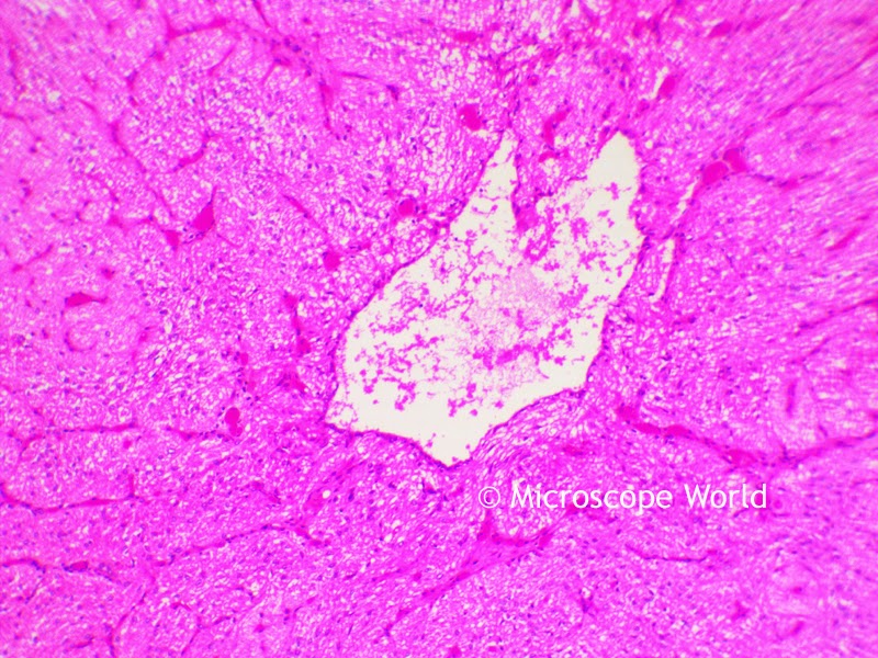
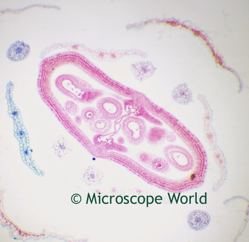
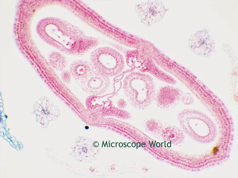
The images below are of a Capsella of a Bud of a flower. These images are of the cotyledon, a significant part of the embryo within the seed of a plant. Upon germination, the cotyledon may become the embryonic first leaves of a seedling. The images were captured using the Richter Optica UX-1D Plan Achromat digital high school microscope.

Microscopy flower image
Capsella of Bud captured at 40x magnification under the UX-1D microscope.

Microscopy image of flower captured at 100x.
Capsella of Bud captured at 100x magnification under the UX-1D microscope.

Capsella bud captured at 400x under the microscope.
Capsella of Bud captured at 400x magnification under the UX-1D microscope.

Dermatology Microscopes

$
0
0
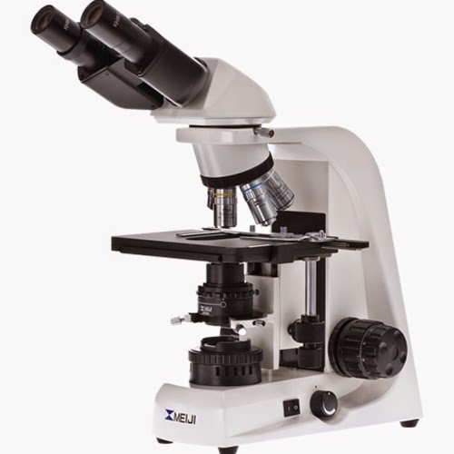
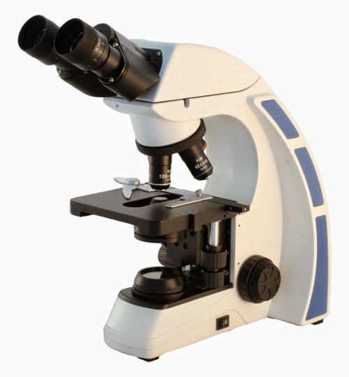
Dermatology microscopes are biological microscopes that require a magnification in the lower range for viewing thin slices of skin samples.

dermatology microscope for professional use
Dermatology Microscope
The MT4000D dermatology microscope shown above includes the following for dermatologists:
  • Plan Apochromat 1.25x, 4x, 10x, 20x, 40x objective lenses.
  • Anti-fungal coating on microscope frame.
  • Low-position mechanical stage for ease of maneuvering slides.
  • Long-lasting LED light with intensity control.
  • Magnification of 12.5x, 40x, 100x, 200x and 400x.

Mustard Seed under the Microscope

Histology Microscopes & Pathology Microscopes

$
0
0
Histology microscopes are used to study cells and tissue in both plants and humans. Pathology microscopes are used to study diseases in human tissues, cells and bodily fluids. Histology and pathology microscopes require a precision setup for viewing thing slices of tissue. An ergonomic head is usually ideal since comfort is important for viewing many slides throughout the day.

Ergonomic pathology and histology microscope.
RB40 Trinocular histology and pathology microscope with ergonomic head.
Some features commonly found on histology and pathology microscopes include:
  • Anti-fungal coating on microscope stage and frame.
  • Magnification ranging from 125x - 400x.
  • Ergonomic viewing head for comfort.
  • Mechanical stage with clinical specimen slide holder.
  • Light focusing on rheostat and condenser.

Small Intestine Microscopy Images

$
0
0
In the small intestine of the human body a network of capillaries transport nutrients including amino acids and carbohydrates. The images below were captured with a high power compound digital microscope.

Microscopy image of small intestine capillaries at 40x.
Small intestine capillaries captured at 40x under a compound microscope.

Capillaries under the microscope at 100x.
Small intestine capillaries captured at 100x under a compound microscope.

Small intestine capillaries under the microscope at 400x.
Small intestine capillaries captured at 400x under a compound microscope.

The microscope prepared slide of small intestine capillaries can be found in the Histology Microscope Prepared Slide Kit from Microscope World.

Hematology Microscopes

$
0
0
Hematology microscopes are used to view blood cells, bone marrow, blood proteins and hemoglobin in order to diagnose, treat, and prevent diseases related to the blood.

Hematology microscopes for viewing blood and bone marrow.
Hematology Microscope
Hematology microscopes are typically coated with an anti-fungal material and often have an ergonomic head as shown above. Magnification of 40x, 100x, 400x and 1000x is used to view blood cells and the blood components. Hematologists deal with diseases such as Malaria, Thalassemia, Leukemia, Clotting Disorders and Anemia.

Image: Pathology Information Educational Resource

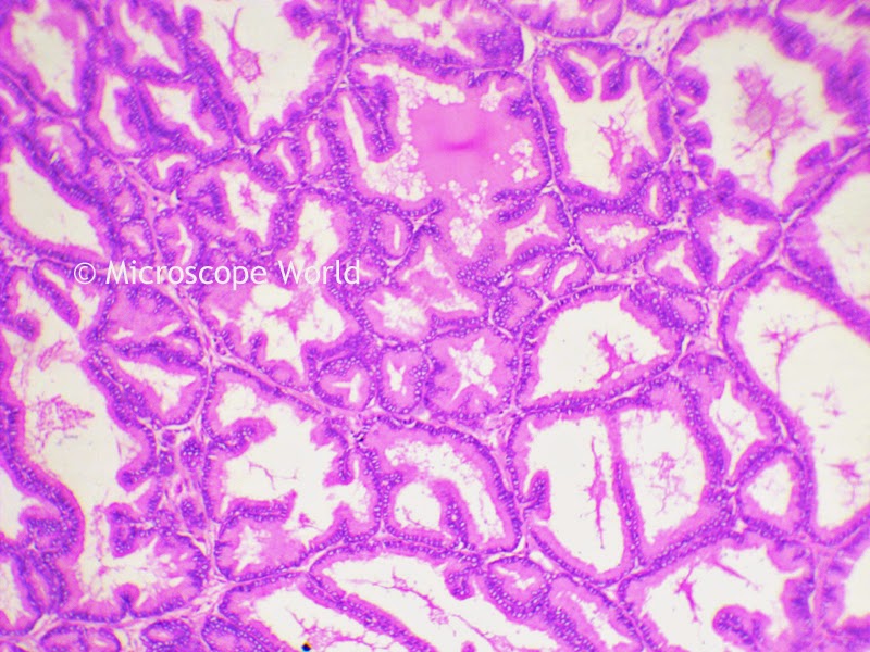
Prostate Gland under Microscope

Stereo Microscope Images

$
0
0
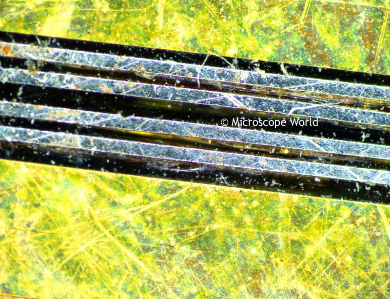
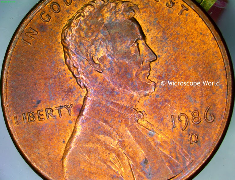
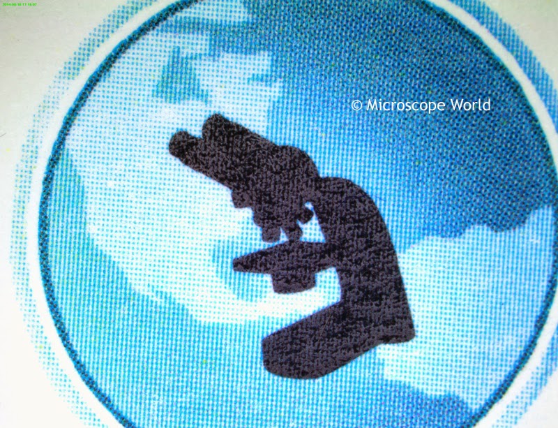
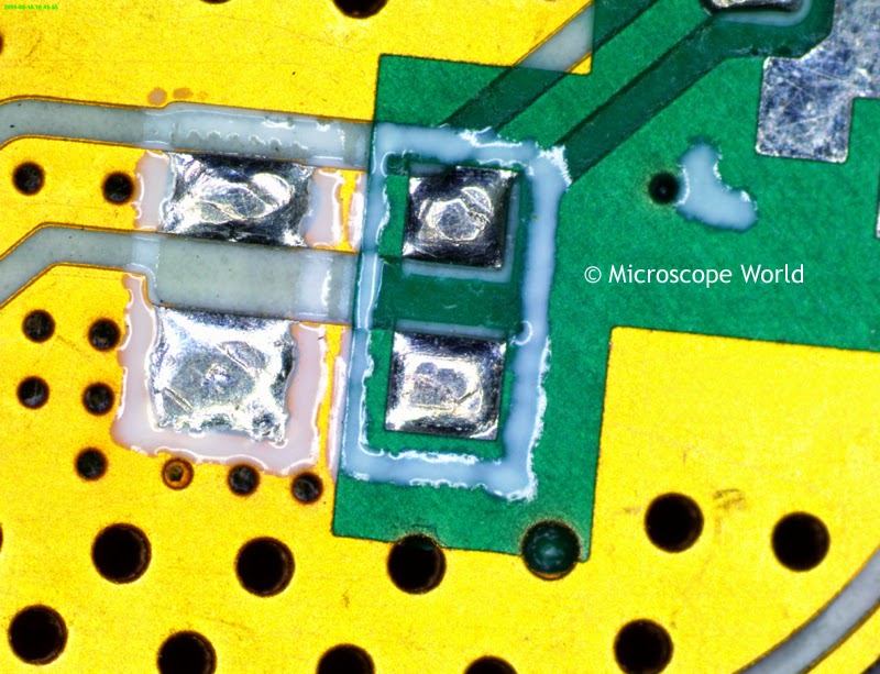
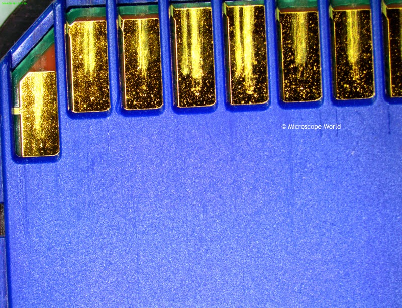
Microscope World just received a new 10 megapixel microscope camera to test out. These are some photos that were captured using the microscopy camera on the S2 dual power stereo microscope.

Printed circuit board under the microscope at 10x.
Printed Circuit Board under the microscope at 10x.

Circuit under the microscope at 10x.

Penny under the microscope at 10x magnification.
Penny under the microscope at 10x.
printed card under microscope
Printed Card Stock under the microscope at 10x.

Printed circuit board under microscope at 30x.
Printed Circuit Board under the microscope at 30x.

microscope image
Industrial Part under the microscope at 30x.

SD memory card captured under the microscope at 10x.
SD Memory Card under the microscope at 10x.


Human Anatomy under the Microscope

$
0
0
In the human body, the pituitary gland is the size of a pea. It is found at the base of the brain. Hormones are secreted from the pituitary gland that help control growth, blood pressure, functions of sex organs, thyroid glands and metabolism as well as some aspects of pregnancy, childbirth, nursing and even temperature regulation and pain relief! For something the size of the pea - it has a lot of important functions!

This is what the pituitary gland looks like under a compound microscope.

Pituitary gland under the microscope at 40x magnification (image).
Pituitary gland under the microscope at 40x magnification.
Gland under the microscope at 100x.
Pituitary gland under the microscope at 100x magnification.

Microscopy image of the pituitary gland at 400x.
Pituitary gland under the microscope at 400x magnification.
The pituitary gland prepared slide is part of the Histology Prepared Slide Kit from Microscope World.

Geologist Microscopes

$
0
0
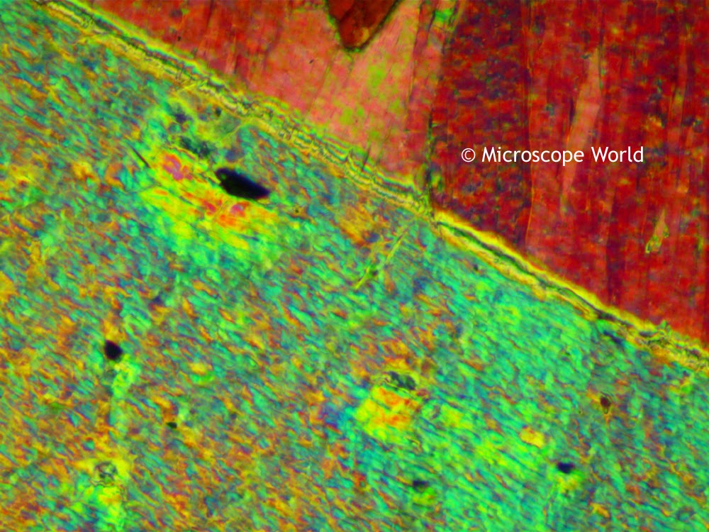
Geology microscopes use polarizing light to examine thin sections of rock. Geologist are looking at the components and features of the rocks along with composition. They take into consideration how the rock was formed and the geological setting of the rock.

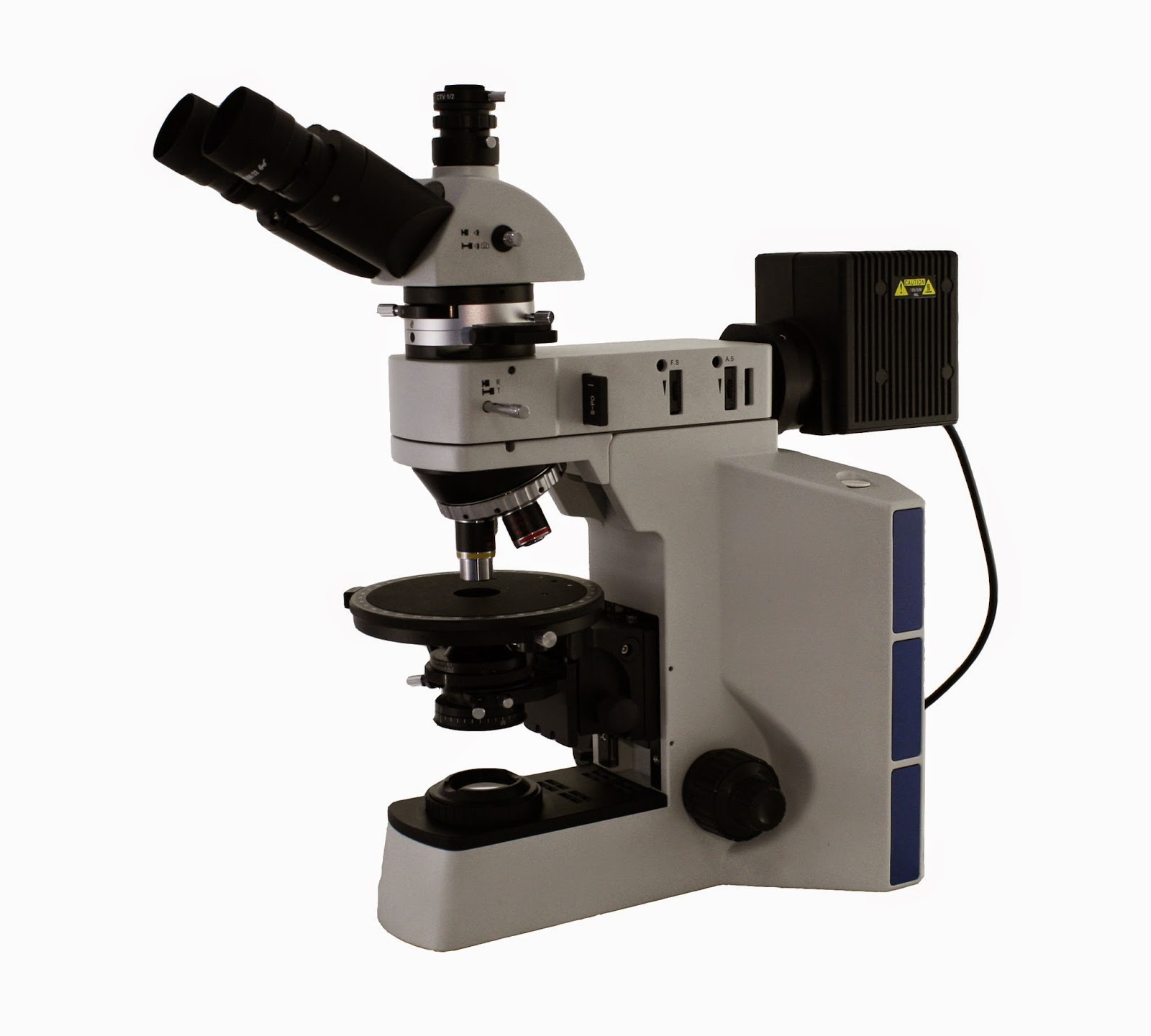
Fein Optic R40POL Geology Polarizing Microscope with reflected and transmitted light.
R40POL Geology Microscope
Geology microscopes are also referred to as petrographic microscopes or polarizing microscopes. These are a few of the features that make up a geologist microscope:
  • Polarizing filter in the light path.
  • Analyzer in the light path between the objective & the eyepiece (works with the polarizer).
  • Circular rotating stage with vernier scales for reading orientations.
  • Bertrand lens allows viewing of conscopic interference patterns.
  • Waveplate to alter the polarization state of the light traveling through it.
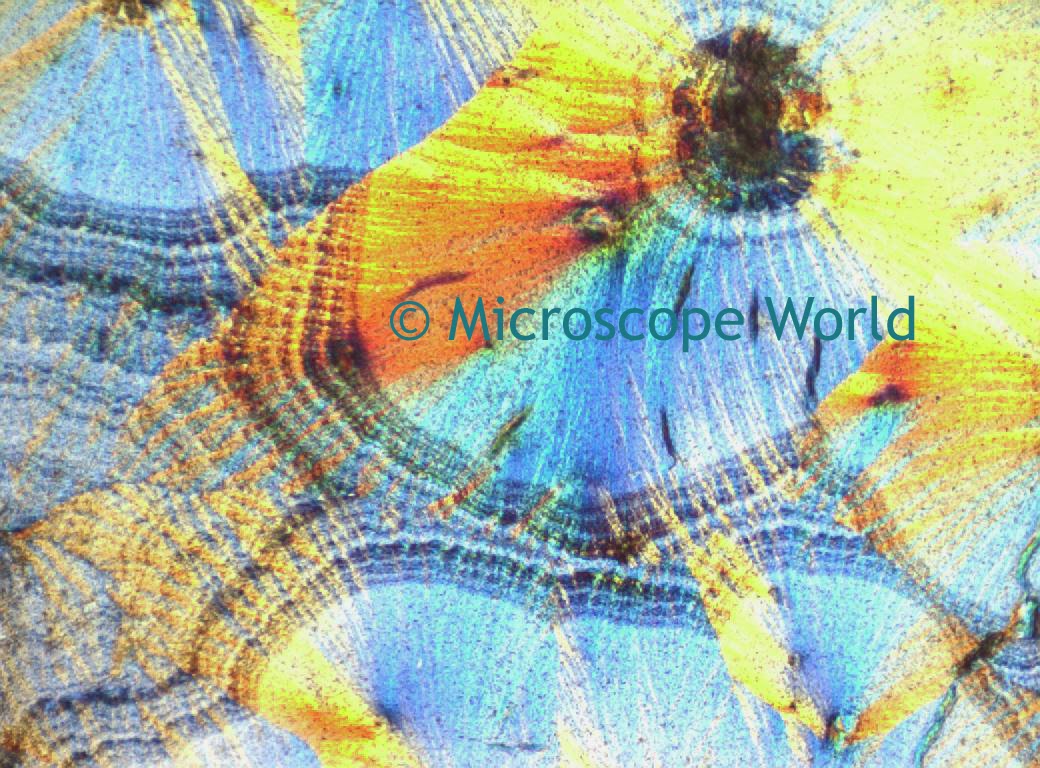
Geology microscope image captured at 200x.
Image captured at 200x under a geologist microscope.
If you have any questions about geology microscopes, feel free to contact Microscope World.

Nerve Fiber Bundles Under the Microscope

Happy St. Patrick's Day!

Swift M10T-MCS Microscopes

$
0
0
Swift Optical has replaced the M10LB microscope with the previously small LCD screen with the new and improved Swift M10T-MCS microscopes. The former small 3" screen has been replaced with a 7" LCD screen.

Swift M10T-MCS tablet WiFi microscopes with 7" screen.
Swift M10T-MCS tablet microscope.
The Swift M10T-MCS is available with semi-plan or plan achromat objectives. There is also a phase contrast version of this microscope - Swift M10T-MCS-MP available.

Some new features of the Swift tablet microscope include:
  • 7" LCD display
  • WiFi, mini HDMI and micro SD card connections
  • 5 mega pixel camera
  • Includes Motic Images software for editing and measuring images.
  • Android operating system.
  • WiFi supporting both hot-spot mode or using an existing WiFi network.
  • Share real-time images with multiple mobile devices.

Diatoms & Vorticella in Standing Water

$
0
0
This water sample was collected from some stagnant pool water and placed under the U2 biological microscope for examination. Notice the diatom and the vorticella on the edge of the algae.

Diatom and Vorticella under the microscope at 400x.
Water from a stagnant pool captured at 400x magnification under the microscope.

Semen Microscope

$
0
0
A semen microscope or sperm microscope is used to identify and count sperm. These microscopes are used when breeding animals or for examining human fertility. You can view sperm at 400x magnification. You do NOT want a microscope that advertises anything above 1000x, it is just empty magnification and is unnecessary.

In order to examine semen with the microscope you will need depression slides, cover slips, and a biological microscope. Below are a few recommend semen microscopes. If you have any questions regarding examining sperm under the microscope feel free to contact Microscope World. Below are a few recommended semen microscopes.

Basic Semen Microscope

Semen microscope
Basic Sperm Microscope
The HS-1M is a basic sperm microscope that goes up to 400x magnification. The LED light is cool and will not heat up or kill any living sperm (in case you wish to view mobility). Coarse and fine focusing make it easy to focus on a crisp and clear image. And the price is affordable!


Intermediate Sperm Microscope

sperm counting microscope
Intermediate Semen Microscope
The UX-1 intermediate semen microscope goes up to 1000x magnification and has Plan Achromat
Objective lenses, offering a crisp fully flat image. Binocular eyepieces make it more comfortable for viewing and eliminate the need to close one eye when looking through the microscope. A mechanical stage makes it easy to maneuver the slide both forward and backward as well as left to right. The microscope has a cool LED light and can be used cordless or plugged in. Both coarse and fine focusing make it easy to focus in on the sperm.


Advanced Semen Microscope

Sperm microscope 1000x
Advanced Semen Microscope
The U2 advanced sperm microscope offers high quality Infinity Corrected Plan Achromat objectives. Magnification of 40x, 100x, 400x and 1000x provides plenty of options for viewing the semen. Binocular viewing head makes the microscope comfortable for extended viewing. Mechanical stage provides ease of slide maneuverability and both coarse and fine focusing make it simple to acquire a crisp and clear image of the sperm. The LED light is cool and lasts a long time.

Tremolite Under a Polarizing Microscope

$
0
0
Tremolite forms by metamorphism of sediments rich in dolomite and quartz. The fibrous form of tremolite is one of the six recognized types of asbestos. This material is toxic and inhaling the fibers can cause cancer. Fibrous tremolite is sometimes found as a contaminant in vermiculite, chrysotile and talc. Tremolite, similar to most minerals, is best viewed under a polarizing microscope.

Tremolite from Switzerland.
Tremolite from Switzerland. Image courtesy: Didier Descouens
All the images below were capturing using the Fein Optic R40POL polarizing microscope with transmitted light.

Tremolite under the polarizing microscope using the 1/4 wave plate.
Tremolite using the 1/4 wave plate under the polarizing microscope, 100x.
Tremolite under the polarizing microscope utilizing the polarizer and analyer.
Tremolite using the polarizer and analyzer under the polarizing microscope, 100x.
Tremolite under a polarizing microscope utilizing the full wave plate.
Tremolite using the full wave plate under the polarizing microscope, 100x.
Polarizing microscope image of tremolite at 100x.
Tremolite under the polarizing microscope, 100x.
Polarizing microscopy image using the 1/4 wave plate.
Tremolite using the 1/4 wave plate under the polarizing microscope, 100x.
Tremolite under a brightfield microscope, 100x (no polarization).
Notice the stark difference between images where polarization was utilized, and the bottom image where only brightfield microscopy was used.

What is a Microscope Reticule?

$
0
0
A microscope reticule (also known as a reticle), is a small piece of glass inserted into the microscope eyepiece for counting or making measurements. This small circular glass disc has an image imprinted on it such as a grid, protractor or ruler. There are a number of different types of microscopy reticules, and each is explained briefly below. Click on the link in order to view a wide variety of reticules in each category.

Ruler Reticules

Microscope ruler reticuleMicroscope ruler reticles are the most popular type of reticule used and are ideal for making basic measurements. Available in both mm and inches, the ruler reticules can be NIST certified for precise measuring that meets certifiable standards.


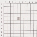
Grid Reticules

Microscopy grid reticle.Microscope grid reticles are frequently used for counting or measuring cells or particles. Measuring particles and cells is made easier, especially if the required task is a pass/fail task. By knowing the size of the grid and whether a particle or cell fits within that grid can make the job of viewing multiple samples at one time more efficient and timely.


Comparator Reticules

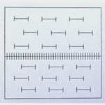
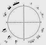
Microscopy comparator reticleMicroscope comparator reticles are typically used for measuring angles. Similar to a protractor, the comparator reticle is available with or without a ruler printed beneath it.


Crossed Scale Reticules

Microscopy crossed scale reticle.Microscope crossed scale reticles are similar to ruler reticles, but have rulers on each of the four axis points. These reticles are useful when needed to measure in several directions from a center point.


Cross Line Reticules

Microscopy cross line reticules.Microscope cross line reticles typically do not have measuring features on them. These microscopy reticles are cross lines, or cross lines with multiple angles, such as a pie.


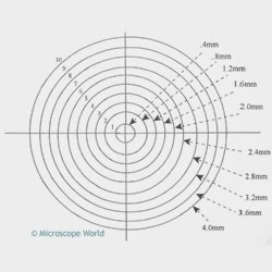
Concentric Circle Reticules

Microscopy concentric circle reticules.Microscope concentric circle reticles are used to measure tolerances from a central point. By using concentric circles, the distance between each circle on the point is a known distance and making measurements of tolerances is much easier.


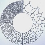
Grain Sizing Reticules

Microscope grain sizing reticle.Microscope grain sizing reticles are used for measuring twine, austenite, carbide, brass, and copper. Additionally grain sizing reticles that meet ASTM E112 grain counting standards, Methods ASTM E-45 A, D and E method, and ASTM C method are reticles that meet specific industry standards.


Whipple Reticules

Microscopy whipple reticles.Microscope whipple reticles have a grid system that usually contains a grid within the main grid. Whipple reticles are intended to enable the measurement of smaller specimen features such as pigment dispersions, colloidal particles, dust, and bacteria.


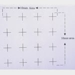
Point Array Reticules

Microscope point array reticles and point cross array reticles.Microscope point dot array and point cross array reticles are used for counting.


Merz Reticules

Microscopy Merz reticles.Microscope Merz Reticles are used to estimate the three dimensional surface areas or the surface density of a component in a given volume, when the component does not have a random orientation. It comprises a test system with parallel curved lines used for measuring the intersection of points.

Pinwheel Reticules

Microscopy pinwheel reticles.Microscope pinwheel reticles are much like the name - a pinwheel with a measuring ruler on each leg of the pinwheel.


Specialized Counting Chamber Reticules

Microscopy counting reticles.Microscope counting reticles include everything from Birnell Scale, Dargie reticle, dirt estimation reticle, Fairs Analysis reticle, Howard mold counting reticle, hole gage reticles, milk smear, Reticulyte counting reticles, Walton & Beckett reticles, Patterson globe reticles, IMA/USP counting reticles, Miller Disc square reticles, Weibel reticles and Porton Counting reticles.

Square Reticules

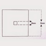
Microscope square reticles.Microscope square reticles and concentric square reticles are commonly used to measure set dimensions.


Once you have your microscope reticle, read this page to find information about how to accurately measure with your reticle.

If you are looking for a particular type of microscope reticle and don't see mention of it on this page, please contact Microscope World, as we carry a number of reticles that are not listed here, as well as custom microscopy reticles.
Art under the Microscope

$
0
0
Linden Gledhill is an artist who has used his education in science and passion for photography to create beautiful microscopy images. Gledhill utilizes a compound biological microscope to capture his images. The microscopy images were captured with a Canon EOS 5D camera mounted on an Olympus BH-2 microscope using 4x, 10x, 20x, 40x and 100x S-Plan Achromat objectives. DIC was used when capturing some of the images.

Microscope setup used to captured images.
Microscopy image of closterium
Closterium (Photo © Linden Gledhill)
Microscope soap film image
Soap Film (Photo © Linden Gledhill)
Food coloring under microscope
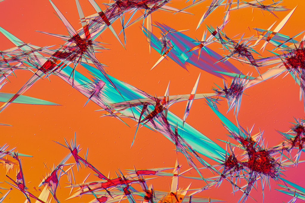
Food Coloring Crystals (Photo © Linden Gledhill)
Diatom under the microscope
Diatom Frustule (Photo © Linden Gledhill)
Microscopy image of algae
Motile Algae, Pandorina Morum (Photo © Linden Gledhill)
Surirella Robusta under the microscope
Surirella Robusta (Photo © Linden Gledhill)
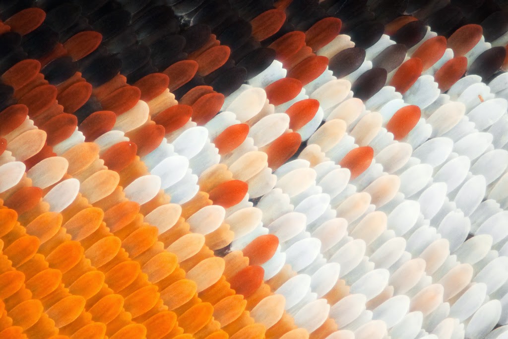
Monarch butterfly wing under the microscope
Monarch Butterfly Wing (Photo © Linden Gledhill)
Microscopy image of a snowflake.
Snowflake (Photo © Linden Gledhill)
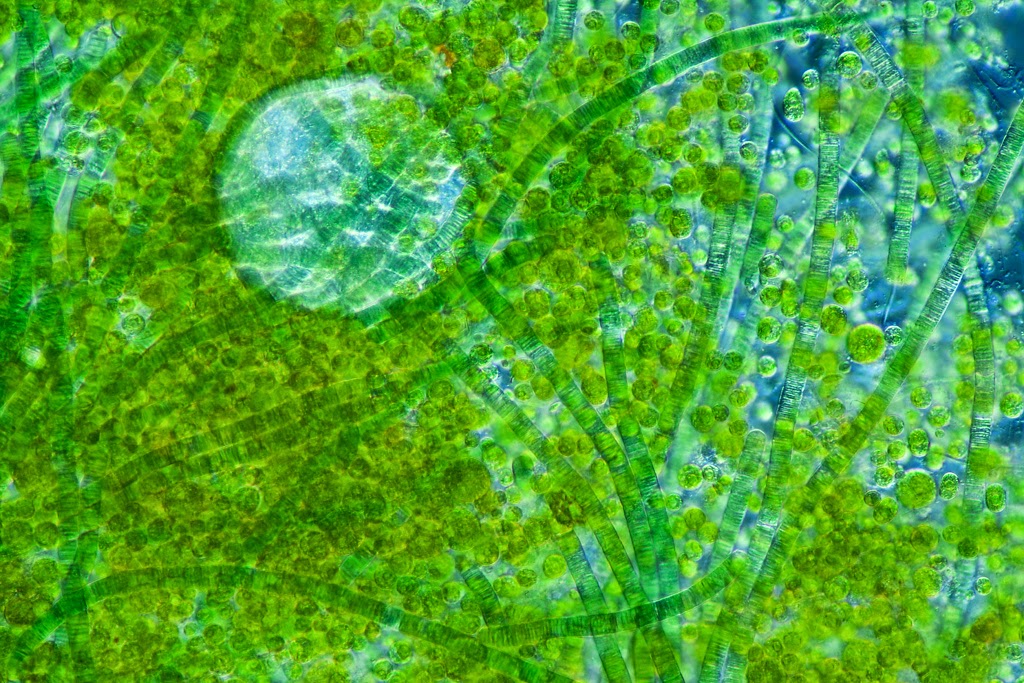
Freshwater algae under the microscope
Freshwater Algae under the microscope (Photo © Linden Gledhill)

Vorticella Under the Microscope

$
0
0
Vorticella Campanula is a fresh water stalked ciliate and usually remains attached to water weeds, other large suspended particles and sometimes to the body of a large zooplankton.

In the video below captured by Ravindra Bakare, Associate Professor of Zoology at Kisan Veer Mahavidyalaya in Wai Dist Satara, Maharashtra, India, stagnant pond water was collected near the University and placed under a biological microscope. Bakare works for the fresh water protozoan diversity around Wai and captured this Vorticella video under the microscope by using a 2 megapixel eyepiece camera at 45x magnification.


Notice that the Vorticella has a ciliary tuft around its peristome and is shown producing a water current for feeding purposes. The unidirectional water current produced by cilia can be clearly seen in the video. The myoneme or stalk is highly contractile when it is attached and covers the area around the radius. 

Thanks to Professor Bakare for sharing this beautiful microscopy work!

ASTM E112 Standard for Grain Sizing

$
0
0
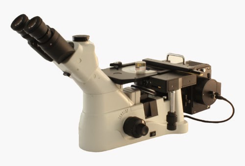
The ASTM E112 standard is commonly practiced in the United States when determining the quality of an alloy. This standard is routinely performed by inspecting a polished sample under a metallurgical microscope to determine grains distribution within a specific area. Typically an inverted metallurgical microscope such as the Fein Optic Mi40 shown below is used.

Inverted Metallurgical microscope Fein Optic Mi40
Fein Optic Mi40 Inverted Metallurgical Microscope
Using this standard, a "G-Number" is derived, which directly relates to the quality of the alloy. The ASTM E112 standard can be performed in one of two ways. Image Analysis software, such as the Infinity Analyze Software included with the Lumenera cameras can be used, or a simple grain sizing reticle can be used.

Grain Sizing Reticle

When using the grain sizing reticle, a polished sample is placed under the microscope and then when looking through the microscope eyepieces the grain sizing reticle is imposed on the image. This reticle allows the technician to estimate the G-Number based on the grain size and how it matches up with the microscope reticle.

Viewing all 422 articles
Browse latest View live