Pathologists study and diagnose disease through examination of organs, tissues and bodily fluids. Digital pathology is the practice of digitizing glass slides and managing the resultant information for later educational, diagnostic, and analytic purposes.
Digital pathology captured images are used for documentation, archiving, teaching, publication and consultation. Microscope digital cameras used in pathology applications use the following features to produce the best results.
High-Fidelity Color Reproduction and Consistency:Pathology is practiced by identifying and acting upon visual cues. In contract to digital radiology, which is confined to grey-scale images, color plays a crucial perceptual role. For this reason, colors captured by the camera should match human perception as much as possible. Additionally color capture should be as consistent as possible from one capture to another.
The fidelity and consistency of color capture is paramount in digital pathology applications. One key consideration is the means by which color wavelengths are filtered prior to being captured by the image sensor. Crucial hardware design choices, such as whether to use mosaicing filters or three different pixel sensors for each RGB color, can affect color quality. In terms of software, reproduction algorithms designed for specific types of pathology must also be carefully designed and tested. Color quality and consistency are also affected by the monitor used to display the resulting image.
Low Noise:Every digital microscope camera suffers from a degree of noise that degrades the image. Due to the stringent quality standards of digital pathology, cameras must exhibit a high signal-to-noise ratio in order to produce images acceptable for medical diagnosis. Camera noise can be divided into fixed-pattern noise or temporal noise. Fixed-pattern noise is produced by variability between pixel to pixel. Using high-quality components and a careful production process can reduce this variability as can proper calibration. Temporal noise is produced during the image capture process. One source is optical (or shot) noise, which is a fundamental and unavoidable property of photos. It is possible to mitigate shot noise through the use of software post-processing algorithms. Another source is electronic noise, produced by the electronic circuitry and semi-conductors during the capture process. Aspects such as size of the photodetector surface, integration time for linear sensors and component quality all play a role.
Wide Dynamic Range / Large Bit Depth:The dynamic range of a camera refers to the range of light intensity that it can capture in one frame. Cameras still struggle to produce low noise images that can match the dynamic range of the human eye. In this specific application, especially when fluorescence specimens are used, it is crucial that both low- and high-intensity signals are captured and displayed to the medical professional.
The fundamental electronic circuitry of an image sensor is one key factor that can significantly impact dynamic range. Another key factor affecting dynamic range is the size of individual pixels. While smaller pixels do increase spatial resolution, they also reduce the number of photos hitting the image sensor, which limits the dynamic range of the resulting image.
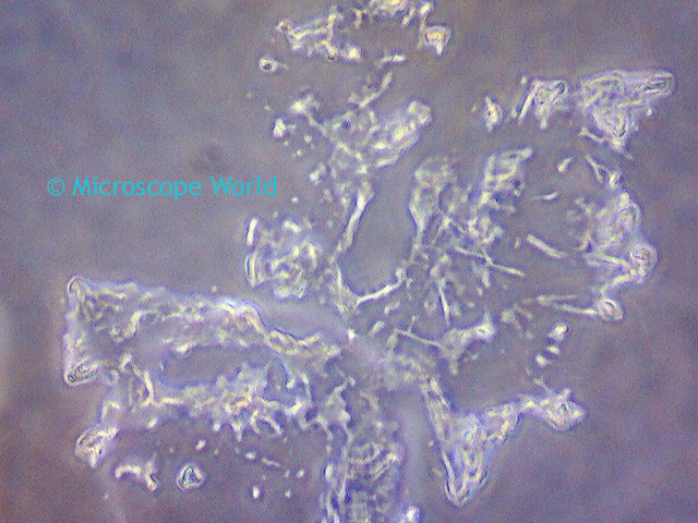
![Infinity 3-3UR microscope camera image of Ilium Brownii Microscopy image captured using Infinity 3-3UR CCD low light camera.]() |
| Ilium Brownii captured with Infinity 3-3UR camera using 10x objective, 10ms exposure, 1.2x gain, 1.0 gamma. |
Excellent Sensitivity:Sensitivity is related to dynamic range and is the lowest light intensity a camera can capture where the amount of noise is still less than the true light signals. Human eyes have lower sensitivity than cameras, which explains why a flash is needed for consumer photography conditions that may seem well-lit to the human eye. High sensitivity is desirable in digital pathology. In particular, fluorescence imaging, with its frequent low-intensity signals, has demanding needs for high-quality images under challenging conditions. Physical components of the camera can affect sensitivity, as can the size of pixels since larger pixels capture more photons.
High Spatial Resolution:A high spatial resolution, meaning the smallest details the imaging system can capture, is a desirable feature of most imaging applications. However, most digital pathology applications, such as certain types of tissue processing, necessitate stringent resolution demands for visual cues and this can push against the theoretical optical resolution limit of visible light.
Large Optical Sensor:The size of the camera sensor affects how much of the microscopy field of view the pathologist can capture at one time.
Fast Frame Rates:Whole-slide imaging requires high frame rates (90 fps or higher) to keep the digital scanning process as fast as possible. In order to match these fast frame rates, the image sensor quality, including many of the factors explained above, must be high enough to perform effectively under these quick conditions.
Standard and High-Speed Data Interface:In order to allow a fast transfer of the image data, the camera must be equipped with a high-speed data interface.
Popular Digital Pathology Microscope Cameras:Quantum efficiency is basically how efficient a camera is at capturing available light, meaning a higher quantum efficiency is desirable. All of the above microscope cameras have a maximum bit depth of 14 bits and output color-accurate raw images.
View all Lumenera microscope cameras here. For more information on digital pathology and setting up your microscope and camera configuration, contact
Microscope World.


