Quantcast
Channel: Microscope World Blog
Viewing all 422 articles
Browse latest View live

Machinist Tool Kits

$
0
0
Microscope World now offers seven machinist tool kits for a variety of tasks including basic inspection, student and apprentice tool kits, depth measurement and routine inspection. Each machinist tool kit is manufactured by Mitutoyo and comes in a mahogany case.

Mitutoyo 64PKA080B basic inspection tool kit from Microscope World.
Basic Inspection Tool Kit
  • 6" Steel Rule
  • Micrometer, Range 0-1"
  • Dial Caliper, Range 0-6"


Mitutoyo 64PKA073B Digital Lite Tool Kit from Microscope World
Digital Lite Tool Kit
  • 6" Steel Rule
  • MyCal Lite Digimatic Caliper 0-6" / 0-150mm
  • Micrometer Lite 0-1" / 0-25.4mm


Mitutoyo 64PKA068A Machinist Caliper and Micrometer Tool Kit from Microscope WorldMachinist Caliper and Micrometer Tool Kit
  • Outisde Micrometer, Range 0-1"
  • Dial Caliper, Range 0-6"
  • 6" Flexible Rule


Mitutoyo 64PKA076B digimatic tool kit from Microscope World.
Digimatic Tool Kit
  • Digimatic Micrometer, Range 0-1" / 0-25.4mm
  • Digimatic Caliper with Absolute Encoder, Range 0-6" / 0-150mm


Mitutoyo 64PKA069A student machinist tool kit from Microscope World.
Student & Machinist Apprentice Tool Kit
  • Outside Micrometer, Range 0-1"
  • 6" Full-Flexible Rule 
  • Test Indicator Set, Range 0.04"
  • Dial Caliper, Range 0-6"


Mitutoyo 64PKA070A depth measurement tool kit from Microscope World.
Depth Measurement Tool Kit
  • Outside Micrometer, Range 0-1"
  • Depth Micrometer with (6 pcs rods), Range 0-6"
  • Full Flexible Rule
  • Dial Caliper, Range 0-6"


Mitutoyo 64PKA071B routine inspection tool kit from Microscope World.
Routine Inspection Tool Kit
  • Outside Micrometer Set (3pcs), Range 0-3"
  • Inside Micrometer (with 6 pcs rods)
  • Combination Set
  • Full-Flexible Rule
  • Test Indicator Set, Range 0.04"
  • Dial Caliper, Range 0-6"
  • Dial Indicator, Range 1.0"
  • Magnetic Stand

Liver Under the Microscope

$
0
0
The liver is a large, meaty organ that sits on the right side of the belly, protected by the ribs. The liver weighs about three pounds and is reddish-brown in color. If you were to touch the liver it would feel rubbery.

There are two large sections that make up the liver: the right and left lobes. The gallbladder sits under the liver, along with parts of the pancreas and intestines. The liver and these organs work together to digest, absorb and process food.

The liver's primary job is to filter blood coming from the digestive tract, before passing it along to the rest of the body. The liver also detoxifies chemicals and metabolizes drugs. As it does so, the liver secretes bile that ends up back in the intestines. The liver also makes proteins that are important for blood clotting and other functions.

The images below are of a pig's liver and were captured under the RB30 biological microscope using the HDCAM4 high definition microscopy camera.

Microscope World image of a pig's liver under a biological microscope at 40x magnification.
Pig's Liver under the Microscope at 40x.

Microscope World image of a pig's liver captured under the microscope at 100x.
Pig's Liver under the Microscope at 100x.

Microscopy image of pig's liver captured by Microscope World at 400x magnification.
Pig's Liver under the Microscope at 400x.

New National Optical D-ELS-1 Stereo Microscope

Textile Exam Microscope for Methods AATCC 20 and 20A

$
0
0
The Association of Textile, Apparel, and Materials Professionals has standards that are required for different test methods. There are several test methods that require the use of a microscope for examining textiles.
  • Test Method AATCC 20 is a qualitative fiber analysis test.
  • Test Method AATCC 20A is a quantitative fiber analysis test.
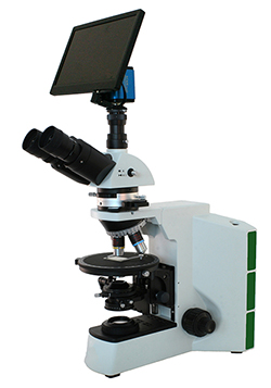
Microscope World has a specific microscope that meets test method AATCC 20 and AATCC 20A standards for textile examination.

Textile Exam Micrsocope for Method AATCC 20 and AATCC 20A
Textile Exam Microscope
The AATCC 20 and AATCC 20A textile examination microscope includes the following:
  • Polarizer and Analyzer
  • Gypsum Full λ Wave Plate
  • Mica 1/4 λ Wave Plate
  • Infinity Corrected Optics
  • Stress Free Plan Achromat 4x, 10x, 40x, 60x Objective Lenses
  • High Definition Camera with Image Capture to SD Card
  • 12" HD Monitor that Connects to Camera
  • Mechanical Stage
Learn more about the AATCC 20 and AATCC 20A textile exam microscope here.

      Microscope Service

      $
      0
      0
      Microscope World provides microscope service and microscope maintenance in Southern California. With over 30 years of technical experience, your microscope will be in good hands whether you need routine maintenance or need a specific problem fixed. You rely on your microscope to perform on a daily basis and if a problem should arise Microscope World understands the number one priority is to get your instrument back up and functioning properly in a timely manner.

      • Parts available for most microscope brands.
      • Volume discounting.
      • Over 30 years of technical experience.
      • Access to parts that are no longer supported by manufacturers.
      • Certified instrument calibrations.
      • Factory trained technicians.
      Learn more about Microscope World's microscope servicing here.

      Images captured with HS2 High School Microscope

      $
      0
      0
      HS-2B Binocular Microscope
      The Richter Optica HS2 is available as a monocular microscope (HS-2M) or as a binocular microscope (HS-2B). The microscope comes standard with 4x, 10x, and 40x Achromat objective lenses and an optional 100x lens is available on some models. This microscope is used most commonly in high schools.

      The images below were all captured using the 40x achromat objective lens that comes on the HS-2 microscopes. A 3 megapixel microscope camera was used to capture the images.


      Microscope World image of Tilia under the microscope at 400x.
      2-Year Tilia captured at 400x with the HS2 High School Microscope

      Microscope World image of monocot and dicot at 400x.
      Monocot & Dicot captured at 400x with the HS2 High School Microscope

      Microscope World image of plant cells captured at 400x magnification under a high school microscope.
      Plant Cells captured at 400x with the HS2 High School Microscope

      Contact Microscope World for more information on high school microscope options.

      Intestine under the Microscope

      $
      0
      0
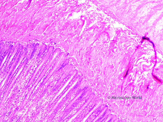
      The Jejunum is the middle portion of the small intestine, connecting the duodenum and the ileum. It is partially responsible for absorbing nutrients into the bloodstream. It is lined with finger-like projections, that are called villi, that move nutrients, vitamins and minerals from the intestine into the bloodstream where they can be used by the entire body.

      The images below were captured with the RB30 lab microscope using a high definition HD microscopy camera.

      Microscope World image of the jejunum captured under the microscope at 40x.
      Jejunum under the microscope at 40x.

      Microscope World image of the small intestine captured at 100x under the microscope.
      Jejunum under the microscope at 100x.

      Microscope World image of the Jejunum (small intestine) under the microscope at 400x.
      Jejunum under the microscope at 400x.

      Contact Microscope World with questions about digital microscopy.

      Museum Display Microscope

      $
      0
      0
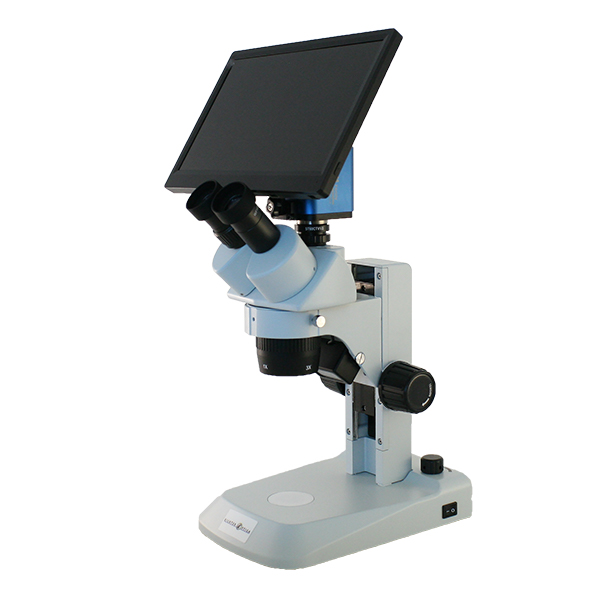
      The museum demonstration high definition display microscope was designed for use in museum and art exhibits. The stereo microscope provides dual magnification of 10x and 30x, ideal for viewing archeological samples, botany samples, fossils and insects. The microscope is equipped with a high definition HD camera and a 12" HD monitor.

      The high definition monitor can face any direction, providing some options for exhibit setup. If the monitor is facing away from the eyepieces the microscope can be set up allowing visitors to look through the eyepieces on one side of the exhibit, while on the other side guests can gather around the monitor and view the samples.

      Alternatively, the museum microscope could be set up in an area where visitors don't have the ability to touch the microscope, but are still able to view the samples underneath the microscope.

      The display microscope has top and bottom LED illumination. This light is a cool light and will not heat up or harm any living specimens. Each light has its own rheostat intensity control.

      Museum display microscope with HD digital camera.
      Museum Display HD Microscope


      Worms found in Cows under the Microscope

      $
      0
      0
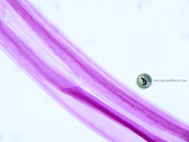
      Bunostomum phlebotomum is a species of worms (nematodes) that are found in the small intestine of calves. The worms are stout and measure 1-3cm in length. Young animals are most commonly affected and only several hundred worms can cause death. Adults often carry worms but do not show any symptoms.

      Diarrhea is the primary symptom in the cows along with signs of anorexia, lethargy and weight loss. Hypoproteinaemia, anaemia and dehydration often occur. Skin involvement due to larvae penetration appears as lesions especially on the feet and limbs. Analysis of fecal samples under a microscope enables eggs to be found, or adult worms can be visualized in the small intestine when an autopsy is performed.

      Veterinarians and ranchers can use Anthelmintics (antiparasitic drugs) for effective treatment of the infection. Preventative antiparasitic treatment may help prevent the disease as well as pasture management to avoid egg and larvae accumulation.

      The image below is Bunostomum phlebotomum that was taken from a calf in Texas. Images were captured using a 3 megapixel microscopy camera on the U2 Lab Microscope.

      Microscope World image of bunostomum phlebotomum under the microscope at 100x.
      Bunostomum Phlebotomum (worm) taken from a cow's intestine, examined under the U2 Microscope at 100x.

      Microscope World image of Bunostomum phlebotomum under the microscope at 400x.
      Bunostomum Phlebotomum (worm) taken from a cow's intestine, examined under the U2 Microscope at 400x.

      Tonsils under the Microscope

      $
      0
      0
      The palatine tonsils are two lymphatic masses, located at the back of the throat on each side of the oral pharynx. The tonsils play a role in protecting the body against respiratory and gastrointestinal infections.

      Each tonsil consists of a network of crypts (pits) that store cells used to fight infection. The tonsils contain B cells, a type of white blood cell that fights infections. They also produce antibodies against polio, streptococcal pneumonia, influenza, and numerous other infections. Antibodies are proteins that help the body identify and attack harmful invaders.

      The tonsils also contain several types of T cells, which are white blood cells that destroy cells infected with viruses and help the body build immunity to infectious organisms.

      The images below are cross sections of Palatine Tonsils that were captured using a biological lab microscope and a microscopy camera.

      Microscopy image of tonsil under the microscope at 40x captured by Microscope World.
      Palatine tonsil under the compound microscope at 40x.

      Microscope World image of palatine tonsil cross section under the microscope at 100x.
      Palatine tonsil under the compound microscope at 100x.

      Microscope World image of palatine tonsil cs captured at 400x.
      Palatine tonsil under the compound microscope at 400x.

      Contact Microscope World with any questions about microscopy or capturing digital images under the microscope.

      Hair Under the Microscope

      $
      0
      0
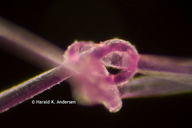
      Harald K. Andersen of Steinberg, Norway captured these images of hair under the microscope. The first image is of a single strand of hair captured through the microscope at 20x magnification. The hair was dyed for the project.

      Hair captured under the microscope.
      (C) Harald K. Andersen. Single strand of hair under the microscope.

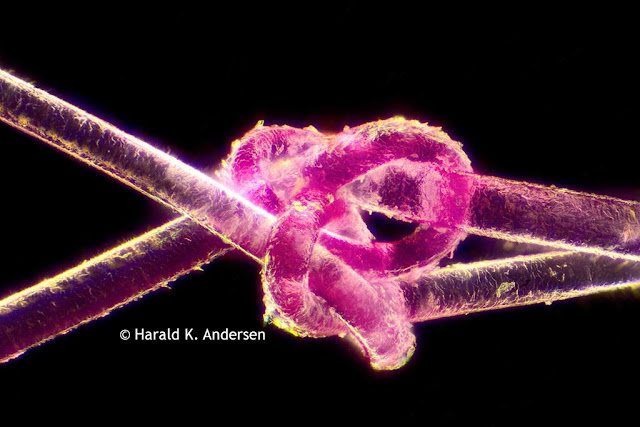
      In order to capture the next image Andersen stacked together 480 images using a software called Zerene Stacker.

      Hair captured under the microscope.
      (C) Harald K. Andersen. 480 stacked images of a single strand of hair under the microscope.

      Andersen mentioned that the hardest part of the project was tying the knot in the hair! A big thanks for sharing the images with Microscope World!

      Mouse Intestine under the Microscope

      $
      0
      0
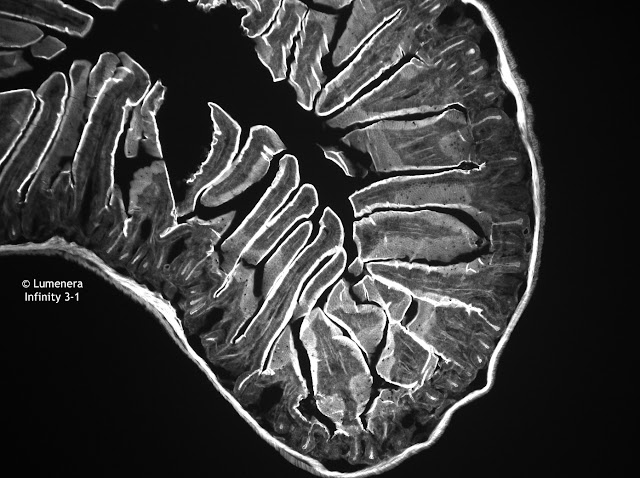
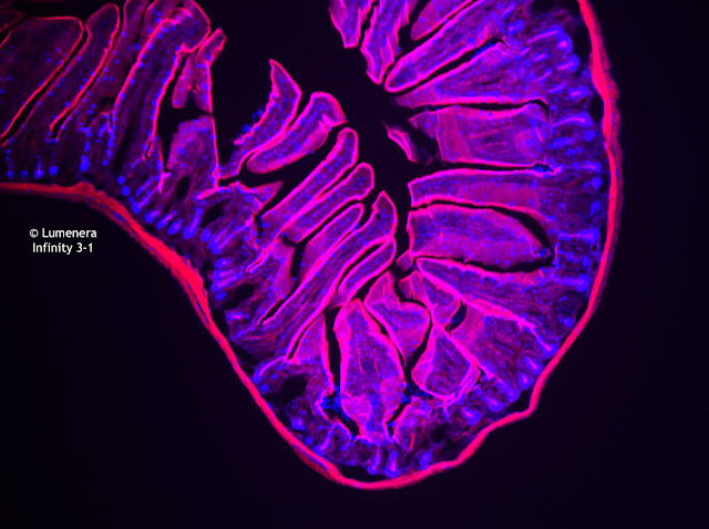
      The images below of a mouse intestine were captured using a Lumenera Infinity 3-1 microscope camera. This is a cooled, CCD camera that is great for low-light situations or where a high dynamic range is required. This 1.4 megapixel camera is ideal for the following applications:
      • Fluorescent Microscopy
      • Green Fluorescent Protein Applications
      • Fluorescent In Situ Hybridization
      • DNA Analysis
      • Live Cell Imaging
      • Brightfield, Darkfield, DIC/Phase Contrast
      • Near IR Applications
      • Histology, Pathology and Cytology
      • Forensic Analysis
      • Semiconductor Inspection
      • Metallurgical Microscopy 
      Mouse intestine under the microscope captured with Lumenera Infinity 3-1 CCD camera.
      Mouse intestine captured with the Monochrome Infinity 3-1 microscopy camera.

      Mouse intestine captured with the Color Infinity 3-1 microscopy camera.
      Mouse intestine captured with the Color Infinity 3-1 microscopy camera.

      Infinity 3-1 microscopy camera image of mouse intestine under the microscope using fluorescence.
      Mouse intestine captured with the Color Infinity 3-1 microscopy camera.

      Contact Microscope World for more information on microscopy cameras.

      High School Binocular Microscope

      $
      0
      0
      The Richter Optica HS-2B is a versatile high school compound microscope. A few features make this microscope one of the most popular microscopes across high schools in the United States.
      • Binocular eyetubes are adjustable from 55-75mm with WF10x eyepieces.
      • 40x, 100x, 400x, 100x magnification.
      • Retractable 40x and 100x (oil) objective lenses avoid damage if moved into contact with the stage.
      • Frame provides easy carrying handle.
      • Cord is attached and not removable - no lost cables in classrooms or at home!
      • LED light is cool and provides long-lasting bulb.
      • Mechanical stage is attached and locked on.
      • Coarse and fine focusing allows for a crisp and clear image.
      • 100~240V allows for use in all countries.
      • Microscope is also available in paired down 400x magnification model, HS-2B-3
      • The HS-2D is a digital version of this microscope.
      • The HS-2-WiFi is a WiFi wireless version of the microscope where you can connect up to six tablets or smart phones.
      Richter Optica HS-2B high school compound binocular microscope provides 1000x magnification.
      Richter Optica HS-2B High School Binocular Compound Microscope

      Richter Optica HS-2B binocular compound microscope.
      Richter Optica HS-2B High School Binocular Compound Microscope

      Sickle Cell Anemia under the Microscope

      $
      0
      0
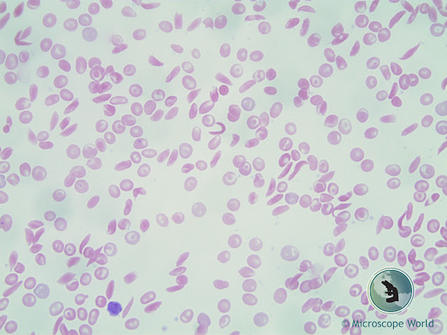
      Sickle cell anemia is an inherited form of anemia. It is a condition in which there are not enough healthy red blood cells to carry adequate oxygen throughout the body.

      Normally, red blood cells are flexible and round, moving easily through blood vessels. When a person has sickle cell anemia, the red blood cells become rigid and sticky and are shaped like sickles or crescent moons. These irregularly shaped cells can get stuck in small blood vessels, which can slow or block blood flow and oxygen to parts of the body.

      Microscopy image of sickle cell anemia at 40x magnification.
      Sickle Cell Anemia under the microscope at 40x.

      Symptoms of sickle cell anemia include fatigue, episodes of pain (when blood cells are being blocked), swelling of hands and feet, delayed growth, and vision problems.

      Microscope World image of sickle cell anemia under the microscope at 100x.
      Sickle Cell Anemia under the microscope at 100x.

      There is no cure for most people with sickle cell anemia. Bone marrow transplant or a stem cell transplant is the only potential treatment, but it is only used in children under age 16 because of the increased risk for older patients. Treatment for sickle cell anemia is usually aimed at avoiding crises, relieving symptoms and preventing complications.

      These images are of sickle cell anemia cells that were captured under a lab microscope using a 5 megapixel microscopy camera.

      Microscope World image of sickle cell anemia captured at 400x.
      Sickle Cell Anemia under the microscope at 400x.

      Learn more about sickle cell anemia here.

      Contact Microscope World with questions regarding microscopes and microscopy cameras.

      Pap Smear Under the Microscope

      $
      0
      0
      A Pap smear is a screening procedure used to test for cervical cancer. The test looks for precancerous or cancerous cells on the cervix. The Pap smear was named after Georgios Papanikolaou, the doctor who determined that the test was a helpful way to detect cervical cancer.

      This is a slide from a Pap smear that was captured under the U2D digital lab microscope at 400x magnification.

      Microscopy image of a pap smear captured at 400x under the U2D microscope.
      Pap smear captured under the U2D Digital Lab Microscope at 400x.

      Contact Microscope World with microscopy questions.

      Tips for Cleaning Microscope Optics

      $
      0
      0
      There are several things to watch out for when cleaning microscope optics. Follow some of these tips when cleaning your microscope lenses to ensure you do not scratch or damage them.

        Tips for cleaning microscope lenses.
      • When cleaning a microscope eyepiece, start out by using a dust blower to remove dust particles.
      • Never wipe lenses with dry tissue as it can scratch the lenses.
      • Do not use any solvents before trying distilled water. A thin film of distilled water can always be created by breathing on the surface of the lens.
      • Do not use abrasive materials such as a dry linen cloth to clean microscope lenses as they may scratch the surface.
      • Older microscopes should not be cleaned using ethanol, diethyl ether or acetone.
      • Use Clean Tip swabs rather than Q-Tips for cleaning.
      • Refrain from using optical spray cans containing pressurized liquid air to clean lenses. These can leave a residue that is difficult to remove.
      • Do not use acids or ammonia to clean microscope objective lenses.
      • Do not try to clean internal optical surfaces, cameras or adapter optics. Contact a microscope service technician if you need to clean these items.

      Contact Microscope World regarding microscope service and repair.

      Motic Panthera Microscopes

      $
      0
      0
      Microscope World is proud to carry the new line of Motic Panthera microscopes. Panthera is Motic's new upright microscope line. There are several Panthera microscopes available.

      Motic Panthera S school microscope with fixed koehler illumination can run off a battery pack if needed.Panthera S School Microscopes

      The Panthera S is a school model. This microscope has Plan SC Achromat objective lenses and an extremely efficient low power illumination that allows the microscope to run on a mobile battery pack for several hours. The Panthera S is a fixed Koehler LED microscope for the educational market and is available in both binocular and trinocular.


      Motic Panthera U Microscope with LightTracer illumination feature.

      Panthera U University Microscopes

      The Panthera U microscopes were created for University use. The Motic Panthera U microscopes have Plan UC Achromat higher quality objective lenses than the Panthera S. The microscope has fixed Koehler 3W LED illumination, and a larger stage than the Panthera S model. The microscope features Motic LightTracer, a digital illumination control. The microscope has a digital intensity knob with coded LED nosepiece that controls the illuminator to offer information on the current light intensity of each objective. Once the microscope is calibrated for each objective lens, the user does not need to adjust the illumination intensity when changing the microscope magnification. The Panthera U is available in binocular or trinocular.

      Motic Panthera C Classic Microscopes

      Panthera C Classic Microscopes

      The Moitc Panthera C microscopes were created for the traditional microscope user. This is a classic all-around microscope that provides both Halogen and LED full Koehler illumination with manual light management. The Panthera C has Plan UC Achromat objective lenses and this microscope includes an integrated USB camera power port (it does not include a camera) and an LED light intensity illumination indicator in the nosepiece. The Panthera C classic microscopes are available in binocular or trinocular.
      Motic Panthera L Life Sciences Digital Microscope

      Panthera L Life Sciences Digital Microscopes

      The Panthera L Life Sciences digital microscopes have built-in digital capabilities. This microscope has Plan UC Achromat objective lenses and Halogen and LED full Koehler illumination. Motic LightTracer provides a digital intensity knob with coded LED nosepiece that controls the illuminator to offer information on the current light intensity of each objective. Once the microscope is calibrated for each objective lens, the user does not need to adjust the illumination intensity when changing the microscope magnification. Digital connection by HDMI, USB, WiFi or RJ-45 allows direct image projection to a monitor or the use of the Panthera App on a mobile device. The Panthera L microscope is available in only one binocular model with a built-in camera.

      Motic Panthera HD Digital Microscope

      Panthera HD Digital Microscope

      The Motic Panthera HD Digital microscope has all the same features as the Panthera L, but it is meant to only be used as a digital microscope. The Panthera HD does not have any eyetubes or eyepieces.


      Contact Microscope World with any questions regarding the Motic Panthera microscopes.

      Motic Images Software Calibration Tutorial

      $
      0
      0
      Motic Images software is included with all Motic microscopy cameras. The software allows for image capture, annotation and one of the most commonly used features of the software: making measurements! However, before you can accurately make measurements with the Motic Images software, it is important to calibrate your microscope with the software first.

      This video walks you through the steps of Motic Images microscopy software calibration.


      Contact Microscope World with questions regarding Motic Images software or Motic microscope cameras.

      Middle School Microscopes

      $
      0
      0
      Microscope World offers several middle school microscopes that are perfect for kids exploring science!

      MDS1 Middle School Microscope

      Richter Optica MDS2 Middle School Microscope is a cordless and rechargeable microscope.
      The Richter Optica MDS1 middle school microscope has several features that make it appealing to middle school classrooms and parents looking for a first microscope for their kids.
      • Cool, LED illumination (the bulb lasts a LONG time)
      • Carrying handle - perfect for younger students
      • Coarse & fine focusing, a must when viewing 400x
      • Cordless & Rechargeable - run the microscope corded or cordless
      • Retractable 40x lens helps teachers protect slides and the lens.

      MDS2 Middle School Microscope

      Richter Optica MDS2 Middle School Microscope has the ability to add a mechanical stage.
      The Richter Optica MDS2 middle school microscope is a step up from the MDS1 in that it allows for the attachment of a mechanical stage. Here are a few features of the MDS2 middle school microscope. (Image shown includes optional mechanical stage).
      • Cool, LED illumination (the bulb lasts a LONG time) 
      • Coarse & fine focusing, a must when viewing 400x
      • Corded microscope
      •  Retractable 40x lens helps teachers protect slides and the lens.
      • Ability to add a mechanical stage, providing more precise control over X-Y movements of slides. 


      MW2-HB4 Middle School Microscope

      MW2-HB4 cordless middle school microscope from Microscope World.
      The MW2-HB4 middle school microscope has a bit of a wider frame, providing added stability to the microscope.  A few features of this middle school microscope include the following:
      • Cool, LED illumination (the bulb lasts a LONG time) 
      • Coarse & fine focusing, a must when viewing 400x
      • Cordless & Rechargeable - run the microscope corded or cordless
      • Retractable 40x lens helps teachers protect slides and the lens.

      Contact Microscope World with questions regarding middle school or student microscopes. 

      Corn Grain Under the Microscope

      $
      0
      0
      Corn is a large grain plant that was first domesticated by people in southern Mexico nearly 10,000 years ago. The ears of corn are actually fruit that produce the kernels (seeds) that people eat. Total production of corn worldwide surpasses that of wheat or rice. However, this is not all used for food. Corn is also used for producing ethanol, animal feed, corn starch and corn syrup.

      The United states produced 361 million tons of corn in 2014, followed by China with 215 million tons and Brazil with 80 tons.

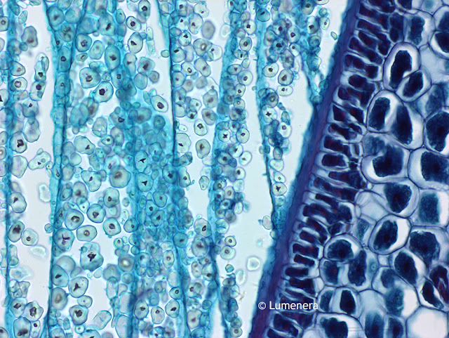
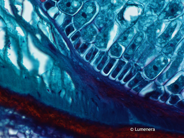
      The images below of a cross section of kernel of corn were captured under the microscope using the Lumenera Infinity 3-3UR microscope camera.

      Microscopy image of kernel of corn.
      Grain of corn captured under the microscope with Infinity 3-3UR microscopy camera.

      Microscope image of a kernel of corn.
      Grain of corn captured under the microscope with Infinity 3-3UR microscopy camera.

      Contact Microscope World for more information on Lumenera cameras or microscopes.
      Viewing all 422 articles
      Browse latest View live