Quantcast
Channel: Microscope World Blog
Viewing all 422 articles
Browse latest View live

Microscope Magnification versus Resolution

$
0
0
In microscopy there are two concepts that many people often think of as a single concept, but they are very different. These two concepts are magnification and resolution. From a technical perspective, resolution is a quantified concept that is defined by the numerical aperture (NA) rating of the objective lenses of the microscope. Numerical Aperture is a number that expresses the ability of a lens to resolve fine detail in an object being observed. Magnification is simply how much an image is enlarged.

Below are two images of the same small printed part with text on it. The first image was captured using a stereo microscope with a lens that has NA of 0.10. This image was captured at 90x. Notice in the image below captured with the stereo microscope it is very hard to even read any of the printed text on the circuit. It should also be noted that it took nearly two hours to capture an image of this quality.
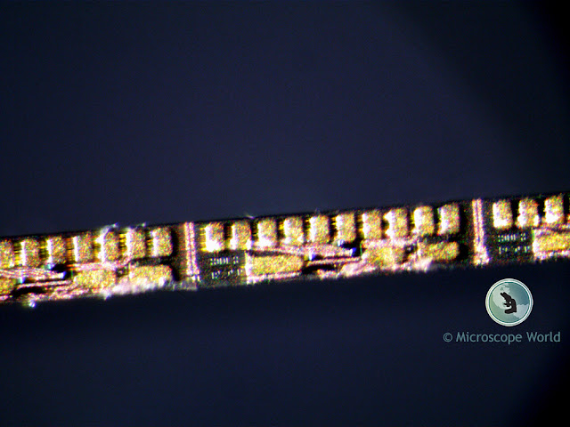
Image of a circuit captured under a stereo microscope at 90x magnification.
Stereo Microscope image captured at 90x, NA 0.10

The next image was captured using a metallurgical microscope with a lens that has NA of 0.30 and a magnification of 100x. This image took a few minutes to capture. The magnifications of the two captured images are similar however, notice how much easier it is to read the printed letters in the image that was captured with the metallurgical microscope. That ability is due to better resolution, which was obtained because of a higher numerical aperture of the lens used.
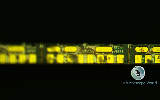
Metallurgical microscope image of a circuit captured at 100x magnification showing high quality resolution.
Metallurgical Microscope image captured at 100x, NA 0.30

Contact Microscope World with questions regarding NA, resolution, magnification or any other microscopy related questions.
Persimmon Endosperm

$
0
0
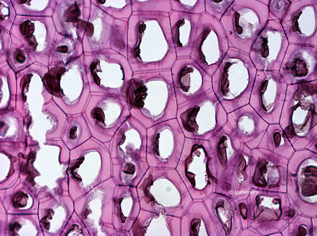
Endosperm is a tissue produced inside seeds following fertilization in most flowering plants. It surrounds the embryo and provides nutrition in the form of starch, though it can also contain oils and protein. This can make endosperm a good source of nutrition in the human diet. The image below is of a persimmon endosperm captured under the microscope using the Lumenera Infinty 2-1 color microscopy camera.

Persimmon endosperm captured under the microscope with Infinity 2-1 microscopy camera.
Persimmon Endosperm under the Microscope captured with Infinity 2-1 Camera

Contact Microscope World with questions regarding microscopes or microscopy cameras.

Microscope Ergonomic Work Station

$
0
0
A proper ergonomic work station when using your microscope can reduce the risk of occupational injuries. UCLA Ergonomics suggests the following tips to maintain a safer environment when using microscopes:
  • Sit in the proper position keeping your back straight and your shoulders back. Distribute your body weight evenly on both hips, and keep your feet flat on the floor.
  • Arrange your work space so that it is close to you.
  • Ensure there is proper padding if leaning on hard surfaces.
  • Work with elbows in close proximity to the body.
  • Work with wrists in a straight and neutral position.
  • Adjust and/or elevate your chair, workbench, or microscope as needed to maintain an upright head position.
  • Adjust microscope eyepieces or mount the microscope on a 30° stand for easier viewing.
  • Repair and clean microscopes regularly. Learn how to clean lenses here.
  • If possible, spread microscope work throughout the day and between several people. 
  • Schedule works breaks. Every 15 minutes, close your eyes or focus on something in the distance. Every 30 minutes get up to stretch and move.
Microscopy is a demanding and rewarding field to work in. By maintaining an ergonomic work space you can ensure years of healthy and pain-free work.

Sources:
"Tips for Microscopy."Laboratory Ergonomics. Ergonomics.ucla.edu, 2012. Web. 27 June 2017.
"Posture for a Healthy Back."Articles. Health.Clevelandclinic.org, 2017. Web. 27 June 2017.

Marmalade Hoverfly under the Microscope Lens

$
0
0
Harald K. Andersen of Steinberg, Norway captured the image below of a marmalade hoverfly. His setup included using the Mitutoyo M Plan Apo 5x Objective lens, connected to his Canon EOS 7D camera.

The marmalade hoverfly (Episyrphus balteatus) is a relatively small hoverfly of the Syrphidae family, widespread through the Palaearctic region which covers Europe, North Asia and North Africa. The upper side of the abdomen is patterned with orange and black bands. These insects often form dense migratory swarms, which may cause panic among people for their resemblance to wasps.

Harald captured this image using a 100 image stack (Canon EF 70-200 f/2.8).

Mitutoyo lens on Canon EOS 7D Camera
Mitutoyo M Plan Apo 5x Objective on Canon EOS 7D

Microscopy image setup for photography
Harald K. Andersen's studio

Harald K. Andersen image of marmalade hoverfly in 100 image stack.
Marmalade hoverfly courtesy of Harald K. Andersen (100 image stack)

Contact Microscope World with questions regarding objective lenses.

Muscle under the Microscope

$
0
0
The human body has 3 types of muscle in it:
  • Skeletal muscle
  • Smooth muscle
  • Cardiac muscle
Skeletal muscle covers the skeleton and gives the body shape. Skeletal muscle is the type of muscle that is most often thought of when the term "muscle" is used.

Muscle under the microscope captured with a PAXcam microscope camera.
Skeletal muscle under the microscope captured with a PAX-cam microscope camera.

Microscopy image of smooth muscle.Smooth muscle (shown at left) is found in walls of hollow organs such as the stomach. Smooth muscle tissue tends to demonstrate greater elasticity than other muscles. It is also found in the urinary tract and digestive tract.


Cardiac muscle is only found in the heart and it is different from the other muscles in the body because it does not get tired. Learn more about the human cardiac muscle here.

Microscopy image of human heart captured at 400x magnification.
Human cardiac muscle captured under the RB30 microscope with a 5mp microscope camera.


Each of the 3 types of muscles in the human body plays an important function in every day life. To learn more about healthy muscles and the roles they play in the human body visit NIH here.

Plant Cell Images from Student Microscope

Chromosomes under the Microscope

$
0
0
A chromosome is a DNA molecule with part or all of the genetic material of an organism. Chromosomes are only visible under a light microscope when the cell is undergoing the metaphase of cell division. During metaphase, when a chromosome is in its most condensed state, the X-shape structure is called a metaphase chromosome.

The images below were captured under the Zeiss Primostar microscope.

Zeiss Primostar microscope images of chromosomes captured at 100x.
Chromosomes captured under the Zeiss Primostar microscope at 100x.

Zeiss Primostar microscope image of chromosomes captured at 400x.
Chromosomes captured under the Zeiss Primostar microscope at 400x.

Contact Microscope World with questions regarding microscopes and different applications.

Lily of the Valley under the Microscope

$
0
0
Convallaria is a genus of flowering plants and includes Lily of the Valley. This is a plant that is sweetly scented and highly poisonous and is native throughout the cool temperate Northern Hemisphere in Asia and Europe.

Convallaria majalis is an herbaceous perennial plant that forms extensive colonies by spreading underground stems and rhizomes.

The images below of Convallaria were captured using the Zeiss Primostar microscope.

Microscopy image of lily of the valley captured under a Zeiss Primostar microscope.
Lily of the Valley rhizome with concentric vascular bundles, captured under Zeiss Primostar microscope at 400x.

Zeiss Primostar microscope image of a lily under the microscope at 100x magnification.
Lily of the Valley rhizome with concentric vascular bundles, captured under Zeiss Primostar microscope at 100x.

Contact Microscope World with questions regarding Zeiss microscopes or capturing images with your microscope.

Understanding Kidney Disease in Dogs

$
0
0
Kidney failure can cause of death in dogs and there are a number of different ways that kidney disease takes shape in dogs. Veterinarians use a microscope to test for some of the following problems in dogs:
  • Glomerular Disease: This is damage to the kidney filters and can be caused by infections such as Lyme disease or cancer.
  • Pyelonephritis: This is an infection of kidney tissues. If a veterinarian finds this infection, the bacteria that is causing the inflammation of kidney tissues can often be killed.
  • Neophrolithiasis: These are kidney stones. Unlike in humans, kidney stones actually do not typically cause dogs too much pain in the early stages.
  • Ureteral Obstruction with Hydronephrosis: This is a kidney blockage. If a dog has kidney stones and they fragment, they can cause a kidney blockage that will not allow urine to pass and the kidneys will swell and become damaged.
  • Tubulointerstitial Disease: This is damage to kidney tabules, often with an unknown cause. This type of kidney disease can only be determined by using a microscope to examine a kidney biopsy (see image below). 
  • Leptospirosis: This is a bacterial infection and can cause kidney disease along with organ problems in both people and dogs. Infection can be caused by contact with urine or other bodily fluids from the infected dog.
  • Toxins: These are medications, household chemicals, and even ordinary food like grapes or raisins. If your pet ingests toxins contact ASPCA Poison Control immediately.
  • Cancer: The good news is that kidney cancer is not common in dogs. The bad news is that treatment options for kidney cancer in dogs is quite limited. If only one kidney is affected it can be removed with a positive outcome. If the cancer is benign or has not spread the outcome can be good as well.
  • Amyloidosis: This is a protein issue with the kidneys where protein deposits replace normal tissue. 
  • Hereditary: Some purebred dogs have a higher rate of kidney disease than others. Additionally, some young dogs will fail to develop normal kidneys.
The image below is of a dog kidney and was captured using a veterinary lab microscope with a microscope camera.

Dog kidney captured under a veterinarian's microscope.
Dog Kidney under the Veterinary Microscope

For more information about veterinarian microscopes contact Microscope World.

Tissue Biopsy under the Microscope

$
0
0
A biopsy is a way for a doctor to diagnose a disease. A sample of tissue or cells is removed from the patient by a doctor and sent to a pathologist to be examined under the microscope.

The following images were captured under a pathology microscope during a tissue biopsy.

Microscopy image of tissue biopsy.
Tissue biopsy captured under a pathology microscope.

Microscopy image of tissue captured under a pathologist microscope.
Tissue biopsy captured under a pathology microscope.

Tissue biopsy captured under a pathologist's microscope.
Tissue biopsy captured under a pathology microscope.

Tissue biopsy captured under a pathologist's microscope.
Tissue biopsy captured under a pathology microscope.

To learn more about different types of biopsies click here. For more information on pathology microscopes contact Microscope World.

Fluorescence Microscopy

$
0
0
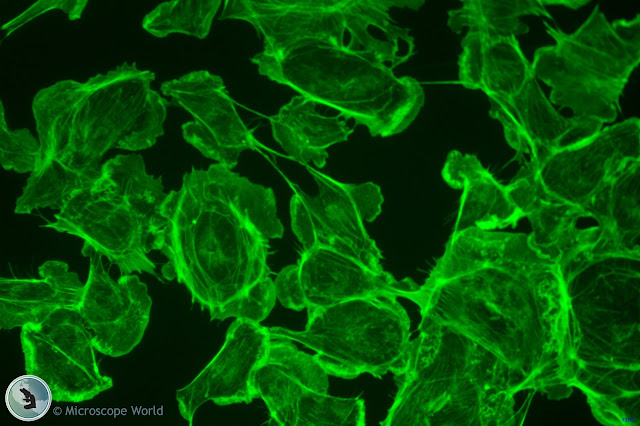
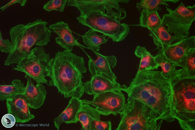
Fluorescence microscopy is an essential tool in biological and biomedical sciences, as well as in material science. The fluorescence images below were captured using a research microscope with a research microscopy camera.

Green fluorescence microscopy image
Green fluorescence image captured with a research microscope.

Monochrome fluorescence microscopy image
Monochrome fluorescence image captured with a research microscope.

Fluorescnece microscopy image showing multi channel fluorescence.
Multi channel fluorescence microscopy image captured with a research microscopy camera.

For more information on fluorescence research microscopes or research microscopy cameras for your specific application contact Microscope World.

Protists under the Microscope

$
0
0
Protists are any member of a group of diverse eukaryotic unicellular microscopic organisms. Protist typically refers to a eukaryote that is not a true animal, plant or fungus because it lacks a multicellular stage.

In order to view protists, a biological microscope is required with 400x magnification.

The image below of a protist was captured using a sample of pond water under the Zeiss Primostar HD digital microscope.

Image of a Protist captured under the Zeiss Primo Star Digital Microscope (c) Microscope World
Protist captured under a digital biological microscope.


Contact Microscope World with microscopy questions.

High School Microscope HSX1

$
0
0
Richter Optica recently introduced the new HSX1 high school microscope. This student microscope has several features that make it appealing to both teachers and students.

Richter Optica HSX1 high school microscope
HSX1 High School Microscope
This high school microscope has a head that rotates 360°, making it easy for students to share the microscope. Teachers like the wider base to help avoid tip-over risk and the locked-on eyepiece. Key microscope features include:
  • Built-in mechanical stage
  • Cool LED illumination
  • Coarse & Fine focusing for crisp images
  • 40x, 100x and 400x magnification
  • Operate corded or cordless
Learn more about the HSX1 microscope here, or contact Microscope World for information about other high school microscope options.
How to Set Up Koehler Microscope Illumination Properly

$
0
0
There are two key areas on a compound biological microscope with Koehler illumination that need to be adjusted in order to set up the Koehler illumination properly: field diaphragm and aperture diaphragm.

The microscope's aperture diaphragm can be found on the condenser of the microscope and it is typically a lever that is adjusted, or a built-in slider on the condenser.

Microscope aperture diaphragm
This is the RB40 microscope, the arrow points to the aperture diaphragm.
The aperture diaphragm on the microscope will usually have numbers on it that correlate to the numerical aperture (N.A.) on your objective lenses.

Microscope condenser aperture diaphragm
Aperture diaphragm on the microscope condenser.

Setting up Koehler Illumination

  1. Start by adjusting the field diaphragm (on the bottom of the microscope where the light is). Focus on your image and open the field diaphragm all the way up so lots of light shines through.
  2. Close the aperture diaphragm (on the condenser) down so you can see the edges of it. Is this dark circle centered in your field of view? If not, use the small centering screws on either side of the condenser to move the circle into the center. 
  3. Open the aperture diaphragm back up until the image contrast is sharp and the edges of the aperture diaphragm are crisp and clear.
This video using the Motic BA410 microscope is a great tutorial to watch if you are uncertain of the steps to take when setting up Koehler illumination on your microscope.


You can learn more about Koehler illumination and setup here.

Spring Flowers under the Microscope


Fluorescence Microscopy

$
0
0
Fluorescence microscopy is a tool used in biology, material science, and biomedical sciences. The fluorescence microscope can reveal the presence of a single molecule. The basic function of a fluorescence microscope is to irradiate the specimen with a desired and specific band of wavelengths, and then to separate the much weaker emitted fluorescence from the excitation light.

The fluorescence microscopy images shown below were captured using the Fein Optic RB30-GFP fluorescence microscope and the Lumenera Infinity 3S-1UR microscope camera.

Fein Optic RB30-GFP fluorescence microscope image.
Fluorescence microscopy image captured using a 60x objective lens.

Fein Optic RB30-GFP fluorescence microscope image from Microscope World.
Fluorescence microscope image, 600x.

Contact Microscope World with questions regarding fluorescence microscopy.

pco Cameras

$
0
0
Microscope World is proud to offer pco scientific cameras. The pco cameras are known for their low readout noise, high frame rates and exposure times that range from nanoseconds to hours. UV and NIR detection and high dynamic range are just some of the pco cameras' unique core competences.

Microscope World offers the pco.panda 4.2 camera.
pco.panda 4.2 Camera


The pco.panda 4.2 camera combines revolutionary sCMOS technology in a compact design. High quantum efficiency (up to 80%) and low readout noise make this camera suitable for countless applications. The USB 3.1 interface enables ultra-speed data transfer and direct power via the USB cable, making external power supplies redundant. Frame rate = 40 fps @ full resolution 2048 x 2048, 80 fps @ 2048 x1024.

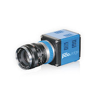
pco.edge camera from Microscope World
The pco.edge 3.1 camera has a sCMOS sensor and is designed for users who require high resolution and high frame rates. This USB3 camera is available in color or monochrome. 50 fps @ full resolution 2048 x 1536, 75 fps @ 1280 x 1024, 160 fps @ 640 x 480.

The pco.edge 4.2 camera has sCMOS technology and can be optionally upgraded with a water cooling system. This camera has high quantum efficiency at up to 82% at peak. 100 fps @ RS fast scan 2048 x 2048, 189 fps @ fast scan 1920 x 1080, 420 fps @ fast scan 640 x 480 when using camera link.

The pco.edge 4.2 LT camera has sCMOS technology and USB3 output. High quantum efficiency of up to 82% at peak. 40 fps @ full resolution 2048 x 2048, 80 fps @ 1280 x 1024, and 170 fps @ 640 x 480.

The pco.edge 5.5 camera has sCMOS technology and can be optionally upgraded with a water cooling system. Quantum efficiency of  > 60% @ peak. With camera link, the frame rates are high: 100 fps @ RS/GR fast scan 2560 x 2160, 201 fps @ fast scan 1920 x 1080, 450 fps @ fast scan 640 x 480.

Woody Stem Under the Microscope

Protozoan Parasite under the Microscope

$
0
0
OE (Ophryocystis Elektroscirrha) is a protozoan parasite that infects butterflies in the Danaus species group, those that host on milkweed. This photo of some Monarch butterfly abdominal scales was taken by Rob Wood with a Canon 80D DSLR through a Richter Optica HS-2 Biological microscope, at 400X. The large objects are scales, while the football-shaped objects are OE spores. Each spore contains 8 protozoa.

Ophryocystis Elektroscirrha captured under the Richter Optica HS-2 microscope.
Ophryocystis Elektroscirrha under the HS-2 microscope.

Thank you Rob for sharing this photo with Microscope World!

Fluorescence Microscopy

Viewing all 422 articles
Browse latest View live