Quantcast
Channel: Microscope World Blog
Viewing all 422 articles
Browse latest View live

Water Silk Algae

$
0
0
Spirogyra is a green algae often referred to as water silk - the less appealing name is "pond scum". Spirogyra is found in fresh water and each cell has two layers - the outer wall is composed of pectin that actually dissolves in water and makes it slimy. The inner wall is composed of cellulose. Chloroplasts are embedded in the cytoplasm and can be seen in the spiral, ribbon-shaped pattern in the image below.

Spirogyra captured at 800x under the microscope.
Pond scum image courtesy J. Jamieson
Image: Australian Biological Resources Study
Spirogyra is a popular green algae to view under a biological microscope due to its ease of sample gathering and its interesting and beautiful spiral patterns.

Glass Beads under Microscope

$
0
0
Owen's Corning manufactures glass beads and they recently needed to measure air bubbles found in some beads.

Microscope World used the HSZ6T-TBL track stand stereo zoom microscope to view the beads. The DCM5.1 5 megapixel microscope camera was used to capture images and the included software was utilized for measuring the air bubbles.

The bead before it was placed under the stereo microscope.

80x magnification (the 2x auxiliary lens was used) with an air bubble that measures 40um.

80x magnification (the 2x auxiliary lens was used) with an air bubble that measures 50um.

 80x magnification (the 2x auxiliary lens was used) with an air bubble that measures 75um.

 40x magnification with an air bubble that measures 145um.

 40x magnification with an air bubble that measures 240um.

If you have a manufactured sample you would like Microscope World to examine under the microscope please contact us.

Microscope Camera Comparison

$
0
0
In order to compare three microscope cameras, Microscope World used the same microscope system and objective lenses to capture all images. The images below were captured using a metallurgical microscope with a 20x, 50x and 100x objective lens. The three microscope cameras used for comparison are shown below and include:
  • 1.3 mega pixel camera (Moticam 1SP with a 1/3" CMOS chip)
  • 3.2 mega pixel camera (DCM3.1 with a 1/2" CMOS chip)
  • 5 mega pixel camera (DMC5.1 with a 1/2.5" CMOS chip)
DCM3.1& DCM5.1 Microscope Cameras

Moticam 1SP Microscope Camera


All images were captured using the HRM300 metallurgical microscope system.

1.3mp Moticam 1SP camera, 20x objective.
3.2mp DCM3.1 camera, 20x objective.
5mp DCM5.1 camera, 20x objective.
The clarity of the images when using the 20x objective (which is producing about 200x total magnification based on the c-mount adapter used), do not vary greatly.

1.3mp Moticam 1SP camera, 50x objective.
3.2mp DCM3.1 camera, 50x objective.
5mp DCM5.1 camera, 50x objective.
As the magnification increases to 50x (about 500x magnification total), the image sensor size becomes more important as far as light is concerned. Notice that the 1.3mp camera starts to look a bit darker than the 3.2mp and 5mp microscope camera images.

1.3mp Moticam 1SP camera, 100x objective.
3.2mp DCM3.1 camera, 100x objective.
5mp DCM5.1 camera, 100x objective.
When using the 100x objective (about 1000x total magnification), the 1.3mp camera produces the darkest image, with less color. The details on this image are still fairly clear. However, the details on the 3.2 and 5mp camera images are a bit more crisp.

Kids Digital Microscope Images

$
0
0
The MW1-LD2 kids digital microscope is the perfect hand-held microscope for kids of all ages. The microscope simply plugs into the USB port on a computer and allows you to view a live image, capture and save images, capture video and edit images.

MW1-LD2 Kids Digital Microscope
The images below of a screw were captured using the MW1-LD2 microscope. The microscope can be held by hand, or with the included stand. Magnification is increased simply by holding the microscope closer to the object.


Microscope Camera Software Multi-Focus

$
0
0
Microscope World carries a line of cameras that include Advanced Panasis Software. One of the features of this microscope software includes the ability to capture two microscopy images and combine them. The manual multi-focus imaging (extended depth of focus tool) allows the combination of several unfocused images into one sharp image. This process is very helpful when the subject height is greater than the available depth of focus, resulting in an image that is partially out of focus. Multiple images captured in sequence from top to bottom are combined into a composite in-focus image.

The microscope cameras that include this advanced software include:
Below image #1 and #2 were combined in the bottom composite image using the microscope camera software. Images were captured using a Huvitz Stereo Microscope.

Circuit Image #1
Circuit Image #2
Combined composite circuit image.

    Lumencor Light Engines

    $
    0
    0
    Microscope World is proud to offer Lumencor light engines for bioanalysis. Lumencor products are made in the USA and the optical excitation subsystems are designed for an array of bioanalytical instruments including fluorescence microscopes. The company has developed innovative, powerful, pure, stable, durable and cost-effective lighting solutions.

    Lumencor Light Engine

    Discrete outputs are available through the UV-Vis-NIR spectrum from a proprietary mix of independently controllable solid state sources. Lumencor’s products provide more power than an arc lamp with the durability, stability, speed and flexibility of a solid state solution.

    The Lumencor light engines are available as a simple light engine package, a computer controllable light engine package, and a computer controllable light engine package with 7 bandwidths.

    Fiber Optic Cables under the Microscope

    $
    0
    0
    Microscope World had a customer that splices and connects fiber optic cables, and they needed to view these parts under a metallurgical microscope. Both the 20x and 50x objectives were used (for 200x and 500x total magnification). The fiber optic cables were viewed with standard brightfield, and with the addition of a green filter.

    200x magnification viewed with brightfield.
    200x magnification viewed with green filter.
    500x magnification viewed with brightfield.
    500x magnification viewed with green filter.

    Flooring Composite Under the Microscope


    Tablets for Microscopy

    $
    0
    0
    Rather than using the conventional microscope camera + laptop computer for capturing images under the microscope, you may want to consider using a microscope tablet camera. The microscope tablet camera is available in both a 7" and 9.7" tablet. The 9.7" camera tablet includes advanced measuring software.

    Boom stand stereo microscope with tablet camera.
    Some benefits and features of using a tablet camera include:
    • 4GB built-in memory.
    • Android tablet with touch screen (can be used for other applications).
    • Built-in c-mount threads that connect to microscope c-mount adapter.
    • Mini SD card slot.
    • Larger screen directly on microscope, rather than having to use cables and place a computer near the microscope system.
    If you are interested in a quote on a tablet microscope system, please email Microscope World.

    Biological Microscope Magnifications

    $
    0
    0
    A basic biological microscope (or laboratory microscope) typically has magnifications of 40x, 100x, and 400x. More advanced compound microscopes will also have 1000x magnification. Any high power microscope that offers magnification above 1000x is providing empty magnification and will not produce a clear resolution image. For magnifications above 1000x a scanning electron microscope is required.

    The images below are frog's blood cells under a biological microscope at each of the three most common magnifications.

    40x magnification

    100x magnification

    400x magnification

    Notice that the nucleus of each cell can be viewed at both 100x and 400x magnification, but at 400x magnification it is much easier to make out.

    A Science Project: Dirt, Bacteria and Soap

    $
    0
    0
    After a long day of playing outside scrape underneath your fingernails and put the dirt (or whatever is under your nail!) on a blank microscope slide. Place the slide under a 
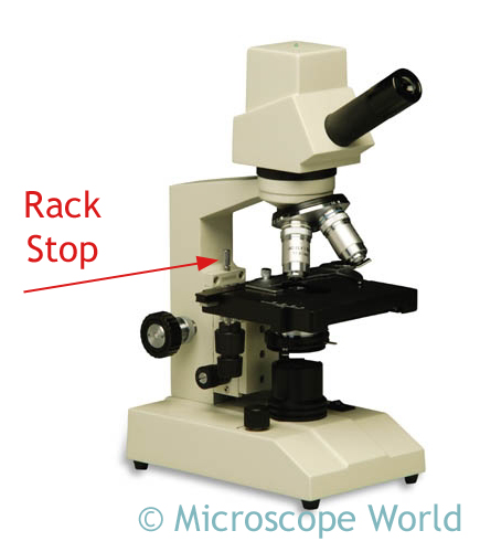
biological microscope. What do you see? Chances are there might be some dirt particles, maybe some carpet fibers and most likely bacteria. If you can, take a picture of what was under your nail.

    Next, take a small scraping of a bar of soap and place it under the microscope. What does the soap look like? Soap is typically made of oils (coconut, palm, olive, etc.), water and Lye (sodium hydroxide). These items are cooked together and when they harden, a bar of soap is formed. The combination of these items breaks down bacteria and dirt that has bonded with the skin when you scrub your hands with water. This washes away bacteria and dirty particles. (You can learn about making your own soap here!)

    Bacteria captured under the microscope at 400x magnification.


    After scrubbing your hands well with soap, take another sample from beneath your finger nail. Can you see any bacteria or particles under the microscope? There should be a lot less particles to look at under the microscope once you wash your hands well, but chances are you will still find some particles to view under the microscope. If there are not fewer particles, you may want to scrub your hands a bit more with soap next time you wash them!

    Watch Microscope

    $
    0
    0
    Viewing small parts on a watch can be tedious on the eyes. By using a stereo microscope with low magnification, viewing the innards of a watch is simple.

    The back plate of this watch was viewed using the MW5-L5 trinocular microscope with a 0.5x auxiliary lens. The auxiliary lens was used both to increase the working distance and also to decrease magnification. Images were captured with the DMC3.1 microscope camera. Total magnification of this captured watch photo was approximately 2.5x.

    Back of watch captured under the stereo microscope.

    Microscope Rack Stop

    $
    0
    0
    What exactly is a microscope rack stop? The microscope rack stop was created to protect the microscope objectives from hitting the stage. It is often easy when looking through the microscope and focusing, to be unaware of how close the microscope objective is to the stage (or glass slide on the stage). Before the microscope rack stop was invented, objectives were sometimes damaged when they hit the stage.

    Rack stop shown on the MW2-HD1 digital kids microscope.
    The rack stop is a small screw that does not allow the stage to move too close to the objective lenses, and keeps the microscope slide at a safe distance from the objectives. The rack stop is set by the factory when the microscope is manufactured.

    It is occasionally necessary to adjust the rack stop. For example, if you were using very thin microscope slides, you might not be able to get the objective lens close enough to your specimen in order to focus properly. In this instance, the rack stop would need to be adjusted so you could focus the microscope and get a clear and crisp image. Adjusting the rack stop simply involves adjusting the screw so it is either higher or lower and will allow the stage to move more or less in one direction.

    If you adjust the rack stop on your microscope, make sure you test the focusing mechanism before looking through the microscope, so that you do not accidentally damage the objective lenses.

    Microscope Disc Diaphragm

    $
    0
    0
    Student compound biological microscopes have either an iris diaphragm or a disc diaphragm beneath the stage. The diaphragm controls the amount of light that is allowed to pass through the microscope stage and up toward the objective lens and ultimately the eyepiece.

    The MW4-H4 biological University microscope has an iris diaphragm that controls the light that passes through the stage. An iris diaphragm has a lever on the side that is simply moved to open or close the iris, allowing more or less light to flood the specimen.

    A disc diaphragm typically has five or six holes of different sizes drilled into a circular disc that is attached beneath the stage. When the disc is rotated, varying amounts of light will pass through the stage.

    The disc diaphragm is used to vary both the intensity and size of the cone of light that is projected upward into the prepared slide. There is no set rule regarding which setting should be used for a particular objective lens magnification. Rather, the setting is a function of the transparency of the specimen on the stage and the amount of contrast required to view the specimen.

    Multi Focus Imaging Software

    $
    0
    0
    Microscope World's most popular software is the Advanced Panasis Software. One of the features of this microscope software includes the ability to capture multiple microscopy images and combine them. The manual multi-focus imaging (extended depth of focus tool) allows the combination of many unfocused images into one sharp image. This process is useful when the subject height is greater than the available depth of focus, resulting in an image that is partially out of focus. Multiple images captured in sequence from top to bottom are combined into a composite in-focus image.

    The images below were captured using a MT7100 metallurgical microscope at 100x magnification. The edge of a flooring sealant material was captured six times, at different focal depths and the microscope software was used to combine them into the final crisp, clear image.

    Image #1
    Image #2
    Image #3
    Image #4
    Image #5
    Image #6
    Composite image combining the above six images using the Multi-Focus feature on the microscope software.

    The microscope cameras that include this advanced software include:

    Reflected Light Microscope

    $
    0
    0
    A reflected light microscope has light that shines down onto the stage from above. There are a few types of microscopes that offer reflected light:
    • Stereo Microscope
    • Metallurgical Microscope
    • Shop Microscope (Field Microscope)
    The Stereo Microscope is the most commonly used type of reflected light microscope. This microscope is used in schools for dissecting, in manufacturing for viewing small parts, and by hobbyists for viewing coins or stamps under the microscope.

    Stereo zoom microscope with 150w halogen fiber optic ring light.
    Metallurgical microscopes offer high magnification, with light that shines down through the objective lens. The metallurgical microscope was created for viewing very small parts of objects that do not allow light to pass through them such as metal, ceramic, stone, etc.

    Huvitz HRM300B-R Reflected Light Metallurgical Microscope   

    These black fibers from a carpet seam were captured using the MT7100 metallurgical microscope.

    A shop microscope (or field microscope) is a small hand-held microscope that can be easily carried out into the field for viewing specimens. These shop microscopes are also frequently used to view fabric during the manufacturing process. The small penlight provides reflected magnification directly onto the object being viewed.

    Shop Microscope
    If you have questions about which type of microscope is best for viewing opaque objects please email us.

    Back to School Science Tips

    $
    0
    0
    It's once again that time of year, new classes, new teachers and an entire year of learning and discovery await kids as they head back to school. Here are a few tips to keep things flowing smoothly at home, especially in the science area.

    1. Use binders for organizing projects or subjects. If you have multiple kids use separate binders for each child. Create a science binder that you will fill up all year long with images and projects. You might want to create sub-folders in your science binder for categories such as Earth Science, Physical Science, Reptiles, etc.
      Photo: Simplify101.com
    2. Create a small science safety kit and put it in a place where your kids know how to locate it should they need to. It doesn't have to be advanced - but if you build the kit with your kids it will make them aware of the importance of science safety.
    3. Create a science storage area where the kids can access supplies when needed, but also know how to put scientific instruments away to protect them. If you have a microscope, store it in a case, or keep the microscope dust cover on it when not in use. 
      Microscope Carrying Case
    4. Many science projects are time-intensive, or require use of space that may need to be used for other activities (for example - the kitchen dinner table). If you have space, set up a small card table that can be used for science projects. Make sure the kids understand the importance of clean up, but also that if their project takes 3 days to complete it is ok to leave the project on that table while it is in-process.
    5. If you can, take time to meet your child's science teacher. Often schools have tools such as microscope cameras or microscope prepared slide kits that kids can use outside of class. There may be items that can't be removed from the science class, but the students might be able to come into the science room for use outside of class hours.
    Make science fun and kids will continue to enjoy the discovery process. Have a great new school year!

    Huvitz Metallurgical Microscopes

    $
    0
    0
    The Huvitz HRM 300 series metallurgical microscopes offer high quality optics with the most advanced model (HRM300BD-RT-3D) including a Z-axis motorized stage and 3D profile imaging software.


    The Huvitz Metallurgical microscopes are manufactured in Korea. The microscopes use the Infinity Corrected Optical System and include Plan Fluor Epi Apochromat objectives that are made for use with brightfield or brightfield and darkfield depending on the microscope purchased.

    metallurgical microscope
    The microscopes can be utilized for brightfield, Nomarski DIC and polarized light. The 100w illuminator ensures that specimens are fully illuminated. Each microscope includes four objectives: 5x, 10x, 20x and 50x. Additional LWD objectives are available. Each microscope includes a 5-year warranty.

    Using the advanced profiler system included in the HRM300BD-RT-3D, the depth of a specimen can be defined and the software will capture image slices that can be compiled into one composite in-focus image, or into a multi-color height profile image

    metallurgical microscope HRM300
    For more information on the Huvitz HRM300 series please email Microscope World.

    Adhesive Under the Microscope

    Most Popular Elementary Student Microscopes

    $
    0
    0
    We are often asked what the most popular first microscopes for kids are, and why. Below you will find a list of the most commonly purchased children's microscope products, and why customers prefer these microscopes.

    1. MW1-L1 Basic 20x Microscope
    • This microscope has no light, which makes it easy to use both indoors and out. There are no cords to trip over and no need to ever replace a light bulb.
    • This microscope is perfect for very small children. Kids can view anything from around the house or out in the yard - leaves, flowers, insects, toy cars, etc.
    child microscope 20x
    MW1-L1 20x Kids Microscope
    • The most popular version of this microscope is the cordless, rechargeable battery version. The microscope can be run off the charger (plugged in), or off the included rechargeable batteries.
    • This microscope is made for viewing slides - kids can make their own (such as viewing cheek cells from inside their mouth), or view prepared slides. At 400x magnification kids will be able to see bacteria, blood cells and protozoans swimming in pond water.
    MW1-HB1 first microscope
    MW1-H1 40x, 100x, 400x Kids Microscope
    • This handheld microscope camera is perfect for children. Rather than taking turns to use the microscope, kids can sit around a computer screen and view the magnified object all together.
    • Magnification varies depending on how close the camera is placed to the object and how large the object is. Camera includes software for capturing images.
    kids microscope camera
    MW1-LD2 Digital Microscope
    • This digital children's microscope offers the same magnification as the kids microscope listed above, but also includes a built-in LCD monitor for viewing a live image.
    • Microscope includes prepared and blank slides for viewing right out of the box.
    MW1-HD2 Digital Children's Microscope

    Viewing all 422 articles
    Browse latest View live