Quantcast
Channel: Microscope World Blog
Viewing all 422 articles
Browse latest View live

Microscopes: Binocular vs. Trinocular

$
0
0

What is the difference between a binocular microscope and a trinocular microscope?

A binocular microscope has two eyepieces for viewing the specimen. Binocular microscopes are more comfortable to look through for extended periods of time than a monocular (single eyepiece) microscope.

Binocluar Microscope
A trinocular microscope has two eyepieces just like the binocular microscope does, but then it also has a third eyetube for connecting a microscope camera.

Trinocular Microscope
When connecting a camera to the microscope, a c-mount adapter is used to connect a microscope camera, or certain point-and-shoot consumer digital cameras can be attached using a microscope camera adapter.

Foam under the Microscope

$
0
0
Foam occasionally needs to be analyzed during the manufacturing process to ensure that the air bubbles in the foam are the appropriate distance apart. These images were captured by Microscope World for a customer that manufactures foam.

Foam captured with a 2mp microscope camera.
On the image of foam above notice the small black ruler imposed on the image. This ruler was created using the basic software that is included with the microscope camera.

Foam captured with a stereo microscope.
The image above was captured using a stereo microscope with transmitted light from beneath the stage. The image was captured with a 2mp microscope camera.

Age Specific Microscope Science Projects

$
0
0
Microscopes provide a great tool for kids to learn about science. Below are some ideas for kids of different ages.

Elementary School Children

Elementary students are often best introduced to science and the microscope with items that are easily found outside such as flowers, insects or leaves. Using a low power simple microscope such as the 20x magnification MW1-L1 or the Little Professor Microscope, these items are easy to view.

Feather captured under the microscope.

Middle School Children

Middle school students are a bit more advanced and enjoy viewing items under the microscope that have more complexity to them. Sugar and salt are perfect items to view under a high power microscope. Have the students look at the granules of sugar or salt on a microscope slide starting at 40x magnification, then moving up to 100x magnification. Once they have viewed the grains of sugar or salt, have them add a drop of water to the sample and notice the changes that take place under the microscope.

Sugar captured at 40x magnification under the microscope.

High School Kids

High school students are learning about things that can't be seen with the naked eye in biology and chemistry. A great science project for high school students is viewing normal tap water versus pond water with a high school biology microscope. The water samples can be placed with an eye dropper on a well depression slide and covered with a glass cover slip.

Bacteria found in pond water.
If you have images you captured using the microscope for a science project we would love to see them. Visit the Microscope World Facebook page and share them with us!

Measuring Edges with a Microscope

$
0
0
Microscope World has a customer that manufactures printed circuit boards.

This is the circuit board the customer manufactures. They needed to measure the very thin edge of the circuit board (when it is flipped on its side).

Using the MC-70 measuring microscope for higher magnification, the circuit board was turned on its side and the images shown below were captured.

Images were captured using a 5.1mp CCD microscope camera.


Stentor (Protozoa) Under the Microscope

$
0
0
Stentor are sometimes referred to as trumpet animalcules because of their horn shape. They typically reach a full grown length of 2mm and are among the largest known unicellular organisms.

Image courtesy Mark Simmons
The image above contains multiple Stentors and was captured using a biological microscope and a consumer digital camera adapter that connected the camera to the microscope.

The body of the Stentor is typically horn-shaped with a ring of prominent cilia around the end of the horn that helps sweep in food and aids in swimming. Stentor are often different colors, including blue. Stentor are found worldwide in freshwater lakes and streams and are usually attached to algal filaments.

Image courtesy Mark Simmons

Jewelers Microscopes

$
0
0
Jewelers microscopes typically have a slightly different setup in that they use darkfield in order to better view precious stones and diamonds, and they also offer the correct lower magnification that is best for viewing jewelry.

Gemological microscopes are available in a variety of prices from simple and basic stereo microscopes that offer a darkfield attachment with a gem clamp, to advanced swivel base full featured gemological microscopes.


All the images above were captured with a very basic stereo microscope and a microscope camera.

Crossline Reticle under the Microscope

$
0
0
A microscope eyepiece reticle is a small circular glass disc that fits into the eyepiece of the microscope and has a ruler, cross-line or other markings printed on it that are imposed upon the microscope image. Eyepiece reticles are most often used for measuring and counting.

The RETCL cross line reticle was placed under the MC70 measuring microscope simply to inspect the reticle markings. The images below were captured at various magnifications using a 5.1mp CCD microscope camera.

Cross-line reticle at 100x magnification.
Cross-line reticle at 200x magnification.
Cross-line reticle at 500x magnification.
The RETCL cross line reticle has 10mm / 100 divisions on each axis with 100um between lines.

Printed Circuit Board


What are Algae?

$
0
0
Algae are organisms that have evolved over time and have the following four things in common:

  1. Algae are simple organims that do not have vascular tissue. Brown algae is the only exception.
  2. The reproductive structures of algae do not have a protective layer surrounding them.
  3. Algae are photoautotrphic, which means that they produce their own food through photo synthesis (using sunlight, water and CO2). Euglena is an exception to this rule, as they ingest other organisms for food because they do not have chloroplasts.
  4. Similar to plants, algae contains chlorophyll. However, not all algae would be classified as plants.
Algae is best viewed through a high power light microscope (also known as a compound microscope or biological microscope). 

Green algae captured at 40x magnification with the BA310 microscope and a 5mp microscope camera.

Blue green algae captured at 100x magnification with the BA310 microscope and a 5mp microscope camera.

Where are Algae found?

$
0
0
In our previous post, we discussed the four characteristics that make up algae. We will now move on to the algae habitat.

Algae commonly grow in any habitat that is moist or where standing water can be found. This includes fresh water, salt water, ice, air, and even in or on other organisms or substrate. In most areas algae fare better in summer rather than winter.

Algae are often regularly found in communities, living within many other different species of plankton, algae, and zooplankton. These communities can tell a lot about the health of an ecosystem. For example, a community of Euglena (a type of Algae), Scenedesmus (genus of Algae - specifically of the Chlorophyceae) and Selenastrum likely indicates a body of water that is rich in mineral and organic nutrients.

Water may be tested for algae content by placing a sample of water on a well depression slide, covering it with a cover slip and viewing at 40x, 100x, or 400x under a biological microscope.

Image of Selenastrum courtesy of Silken Tent.
Image of Scenedesmus courtesy EPA.

Viruses In the Ocean

$
0
0
Viruses are the smallest and most abundant components of plankton. They range in size from 0.02 – 0.2um in diameter. In one liter of surface seawater you would find up to 10 billion viruses!

When most people think of a virus they associate it with sickness, but viruses are essential to the regulation of saltwater and freshwater ecosystems. Most marine viruses are bacteriophages, infecting the bacteria in aquatic microbial communities, and are harmless to plants and animals.

Virus particles (virions) are not alive in that they have no metabolism, no mobility with which to swim, and they do not grow. Instead, new viruses are assembled within a host cell. In order to replicate, a virus must contact an appropriate host cell and transfer its genetic material to the inside of that cell. Once inside, the viral genome directs the host cell to produce new virions. When these newly created virions exit the host cell (often killing the cell in the process), they are again lifeless particles drifting in the sea, waiting another chance encounter with the right type of cell.

Viruses are so small that they can only be viewed with a scanning electron microscope, or they can also be detected as small dots using epifluorescence microscopy with a sufficiently bright fluorescent DNA stain.

Autotrophs

$
0
0
Autotrophs are organisms that produce complex organic compounds (such as fat, carbohydrates or proteins) from simple substances that are found it the surroundings generally by using light from photosynthesis. These organisms make their own food by "fixing" carbon dioxide - basically they self-feed. Photoautotroph plants fix carbon using energy from the sun. Chemoautotrophs use chemical energy.

On land, most photosynthesis is performed by multicellular life (plants), but in the ocean, nearly all photosynthesis is done by single-celled organisms. The most abundant photosynthetic organism on earth is a prokaryote.

Bacteria is a prokaryote. The image below of bacteria was captured using the digital biological microscope MW4-HD2 with phase contrast.

Bacteria captured under the microscope at 100x magnification.

You can learn more about prokaryotes in the ocean here.

Types of Microscope Plan Objective Lenses

$
0
0
Biological microscope objective lenses are available in several options. Each of these microscope objective lens options are described below.


Achromat Objectives - these are the most basic high power microscope objectives. Typically the outer edge of the circular image seen through the microscope will be slightly out of focus due to the curvature of the lens.

Semi-Plan Achromat Objectives - these are one step up from basic microscope objectives in that the field of view (circular image) has been corrected a bit and the outer edges will be in focus more than the standard achromat lens. However, the entire field of view will not be a completely flat and focused field - the very outer edge will still be a bit out of focus.

Plan Achromat Objectives - these microscope objectives have been corrected for lens curvature and the entire image plane is flat and in focus - including the edges of the image.

Plan Apochromat Objectives - these microscope objectives are also referred to as Plan Apo. These objectives have been corrected for four colors chromatically and spherically. Plan apochromat objective lenses are the best objectives to use for critical resolution and color photomicrography. These objectives typically have a shallower depth of field.

Plan Fluorite Objectives - these microscope objective lenses are corrected for four wavelengths, but not quite as extensively as plan apochromat objectives are. Plan fluorite objectives are well suited for photomicrography and are typically a bit less expensive than plan apochromat objectives.

If you are looking for a specific type of microscope objective lens please email Microscope World for further information.

Kids Microscope Project: Dust

$
0
0
This is a simple and fun microscope project for kids of all ages.

What you need:
Collect the dust with your finger if it is thick enough on a window sill or in a door jam. Another great source for interesting dust to observe is from the vacuum cleaner. An intake vent for air is a great place to collect dust - look at your hair dryer or the fan vent on your computer.

If using a stereo microscope, place a small amount of dust on the glass stage. Try viewing the dust with only the light from beneath the stage, then only the light from above, and finally both lights together. What differences do you notice? Can you find any specific particles such as insect legs or carpet fibers? What about small rocks - can you tell what they are made out of?

If using a biological microscope place a small amount of dust (you may want to use an old toothbrush to do this) on the glass slide and place it beneath the microscope starting at 40x magnification. Once you have the dust particles in focus, move the slide a bit to view other particles. What do you see? Draw images of the particles you see. After you have viewed the dust a bit, you may want to place it on a depression slide with a cover slip and add a small drop of water. How do the particles change when water is added?

If you have captured some images of dust under the microscope we would love to see them posted on the Microscope World Facebook page.

Microscope Objective Abbreviation Guide

$
0
0
Microscope objective lenses (particularly those made for biological microscopes) often have abbreviations inscribed on them that can at times be confusing. The list below may help you decipher exactly what the inscription on your microscope objective lens stands for.
 ABBREVIATION  & Explanation:
  • ACHRO = Achromatic aberration correction
  • EF = Extended field (The field of view is less than plan)
  • PL, PLAN, PLANO = Flat field optical correction
  • N, NPL = Normal field of view plan
  • UPLAN = Universal plan (Brightfield, darkfield, polarized light)
  • LU = Luminous universal (brightfield, darkfield, DIC, polarized light)
  • PLAN APO = Apochromatic and flat field correction
  • FL, FLUOR, FLUAR = Fluorite aberration correction
  • L, LL, LD, LWD = Long working distance
  • ELWD = Extra long working distance
  • SLWD = Super long working distance
  • CORR, CR = Correction collar
  • I, IRIS = Adjustable numerical aperture with iris diaphragm
  • OIL, OEL = Oil Immersion
  • WATER, WI, WASSER = Water Immersion
  • HI = Homogeneous Immersion
  • GLY = Glycerin Immersion
  • DIC, NIC = Differential or Nomarski interference contrast
  • CF, CFI = Chrome-Free or Chrome-Free infinity corrected
  • ICS = Infinity color-corrected system
  • RMS = Royal Microscopical Society objective thread size
  • M25, M32 = Metric 25mm or 32mm objective thread
  • PHASE, PH, PC = Phase contrast
  • PH 1, 2, 3 = Phase condenser annulus 1, 2, 3
  • DL, DLL, DM, BM = For phase contrast: dark low, dark low low, dark medium, dark bright
  • PL, PLL = For phase contrast: positive low, positive low low
  • PM = For phase contrast: positive medium
  • NL, NM, NH = For phase contrast: negative low, negative medium, negative high contrast
  • P, PO, POL, SF = Strain-free, made for polarized light
  • U, UV, Universal = UV transmitting for UV-excited epifluorescence
  • M = Metallographic (no cover slip is used)
  • NC, NCG = No cover slip
  • EPI = Epi illumination
  • TL = Transmitted light
  • BBD, HD, B/D = Bright or dark field (Hell, Dunkel)
  • D, DF = Darkfield
  • H = For use with a heated stage
  • U, UT = For use with a universal stage
  • DI, MI, TI = Interferometry, Noncontact, Multiple Beam (Tolanski)

The Kidney

$
0
0
Kidneys are organs that serve several essential regulatory roles in most mammals. Kidneys regulate electrolytes, they are essential to the urinary system, and they regulate blood pressure as well as filtering the blood to remove wastes.

Image courtesy Gray's Anatomy
The kidneys primary function is to filter wastes out of the body by creating urine. The kidneys also play a role in separating waste from vital nutrients and allowing the body to absorb these vital nutrients.

Kidney image courtesy Fr. Dr. Julia Enrick
The image above of a kidney was captured using a biological microscope and the Jenoptik C5 microscope camera.

Darkfield Illumination in Stereo Microscopes

$
0
0
Darkfield observation in stereo microscopy requires a specialized stand that contains a reflection mirror and light-shielding plate to direct a hollow cone of illumination towards the specimen at oblique angles.

The principal elements of darkfield illumination are the same for both stereo microscopes and more conventional compound (biological) microscopes, which often are equipped with complex multi-lens condenser systems.

One example of a simple add-on darkfield kit for a stereo microscope is shown below. The darkfield kit drops into the stage plate position on the MW5-L5 stereo zoom microscope.

Darkfield on a stereo microscope.
Gemologists often use darkfield for viewing diamonds and precious stones. Gemology microscopes therefore typically have a darkfield feature built into the gem stereo microscope stand.

The GEMZ-5 gemology microscope shown above has an arrow pointing to the built-in darkfield unit on the microscope.

Mossy Algae under a Microscope

$
0
0
If you have ever walked along a river or stream you have probably noticed a mossy green alage on the rocks. If you try to step on the rocks, it is slippery.

Image: W. Bourland
This green, soft algae is referred to as cladophora. Cladophora is also known as "river weed" and in Laos it is even eaten as a delicacy in the form of dry sheets, similar to Japanese nori.

Cladophora is a filamentous bloom, meaning it is an algae with longer tendrils. It is often found on rocks in streams or rivers and growing around waterfalls. When viewed under a high power biological microscope, then longer tendrils have multiple fingers that shoot off of each branch.

Image: W. Bourland

Zoology Student Science Project

$
0
0
Different animals and creatures habitat different areas of the country. You don't need to travel anywhere for this science project - simply start in your own back yard! Then, if you ever travel somewhere for a vacation, you can repeat this science experiment in a new area and compare your findings.

This project is made to classify animals into different groups. How many different groups of animals and creatures are located near your home? Your backyard may be more bio-diverse than you think!
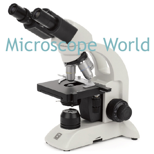
Materials Required:
Microscope for viewing samples
Pen and paper to record findings
Optional - microscope camera to capture images.

The goal of the project is to observe things that move - animals both large and small. If you are able to pick up the smaller insects, take them home to view them under the microscope. Some good locations for observation include:
  • Parks
  • Your back yard at home
  • A creek or pond
  • Wooded areas
  • The desert
  • A garden
Note the time of day you performed your experiment (some animals only come out at night - they are referred to as nocturnal).

 Antennae captured under the microscope at 100x magnification.

In your notebook jot down the following:
Habitat (were you in the grass, digging in the soil, was the animal up in a tree?)
Animal (earthworm, squirrel, ant, snail, etc.)
Draw a sketch or capture an image under the microscope.
What Phyla does this animal belong to? (You may need help from a teacher or a book to determine this - below you will find a few examples to help you.)
  • Arthropods = ants, grasshoppers, spiders
  • Annelids = earthworms
  • Molluscs = snails & slugs
  • Chordates = birds & squirrels
  • Echinoderms = starfish
  • Porifera = sea sponge
Record your findings and share with your class. What category above do you fall into? Which Phylum did you find the most animals for?

Green Algae Oedogonium under the Microscope

$
0
0
When scooped up from a pond or still water source Oedogonium will adhere to your hand, rather than slip through your fingers.

Photo: D. Patterson
Oedogonium is a filamentous green algae with filaments that are only one cell thick. This type of algae is occasionally free-floating, but is often attached to other aquatic plants.

Photo: Curits Clark
Oedogonium thrives in fresh water that is not disturbed.

Photo: D. Patterson
When Oedogonium is viewed under a biological microscope, the single cell filaments are more visible. If this algae becomes out of control in fresh water environments the water is often treated with copper to limit its growth and keep it from invasively overtaking the pond.
Viewing all 422 articles
Browse latest View live