Quantcast
Channel: Microscope World Blog
Viewing all 422 articles
Browse latest View live

Articulated Arm Stereo Zoom Microscope


Fun Things for Kids to View with Microscopes

$
0
0
There are two types of microscopes: high power biological microscopes and low power stereo dissecting microscopes. This post lists fun items for kids to view with a low power stereo microscope.

A stereo dissecting microscope typically will have light both above and below the stage. This type of microscope is perfect for viewing any type of sample that does not allow light to pass through it. Magnification on a stereo microscope is usually between 10x-40x.

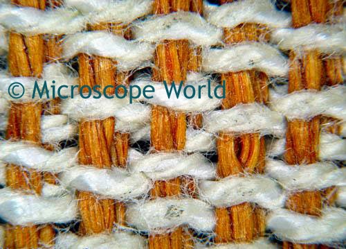
fabric under stereo microscope
Fabrics look very interesting under the microscope!
screw seen under microscope
Hardware under the Microscope
ants under the microscope (image)
Insects (this one is an Ant) under the microscope!
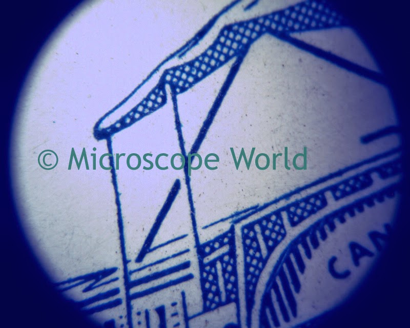
Postage Stamp under stereo microscope
Stamp or newsprint under the microscope

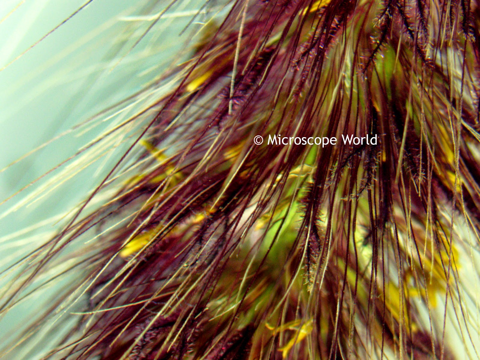
microscope image of cattail
Cattail under the Microscope

flowers under microscope
Flowers under the microscope

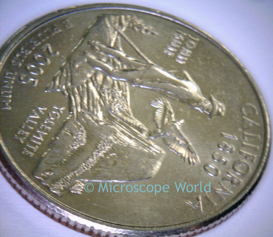
coin image under microscope
Coins under the microscope
Have you captured any interesting images under the microscope? We would love to see them - share them on our Facebook page!

Making Measurements with Microscopy Tablet Cameras

Optic Nerve under the Microscope

$
0
0
The optic nerve transmits visual information from the retina to the brain. The optic nerve's function includes special somatic afferent, which carries the sensory modality of vision.

The images below of the optic nerve were captured using the Richter Optica U2 biological microscope at 400x magnification.

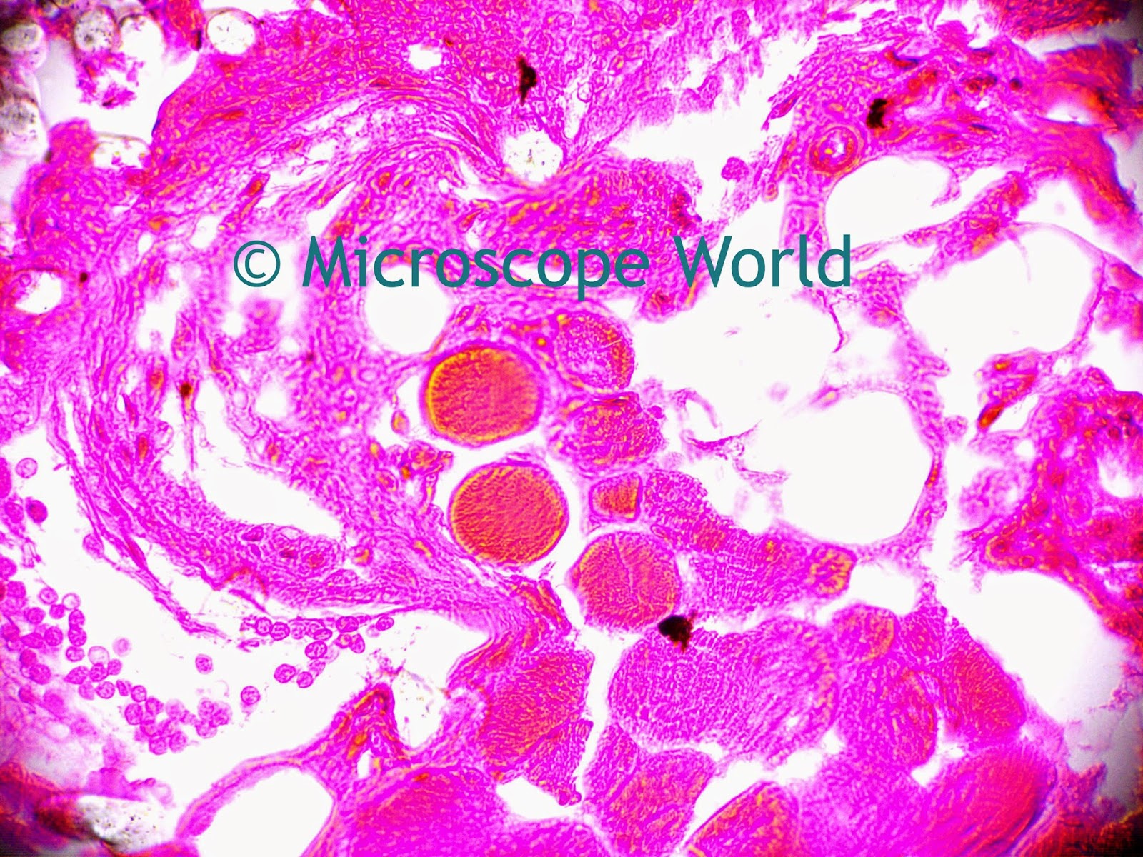
optic nerve under microscope 400x
Optic Nerve under a brightfield microscope.
The images were captured using a 5 mega pixel microscope camera.

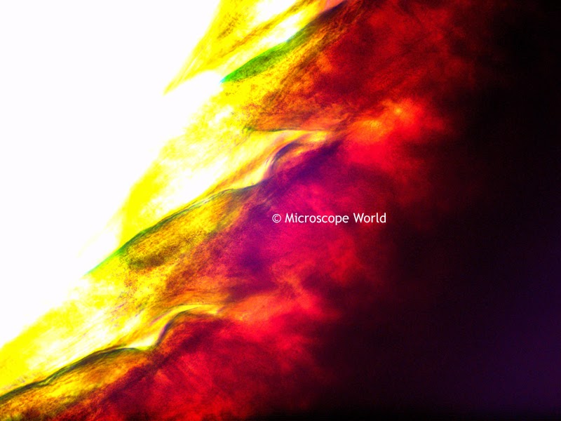
Optic Nerve Epi Fluorescence Microscope Image 400x
Optic Nerve captured under epi fluorescent microscope.
The optic nerve prepared slide can be found in the Histology Musculoskeletal slide kit.

Kids Science Microscope Activity: Yeast

$
0
0
This is a fun science project for kids! You will need the following:
PART I:
  • Place the packet of yeast in the bowl.
  • Add 1 cup warm water and 2 tablespoons of sugar. Mix well.
  • Transfer the liquid to the bottle and place the balloon around the neck of the bottle.
After 10 minutes what to do you notice? Did the balloon start to inflate? This is due to carbon dioxide being released from the yeast.

PART II:
  • Put a drop of the yeast mixture on your microscope slide.
  • Place a cover slip on top and look at the yeast at several magnifications.
  • Draw an image or capture a picture of the yeast at 400x magnification.
  • Can you see the yeast cells?
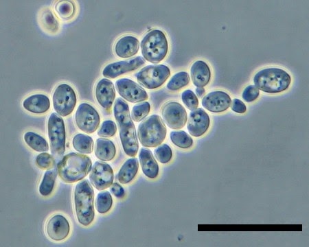
yeast under microscope
Yeast cells in young wine. Image courtesy microbiological garden.
Yeast is used to make bread, wine and beer through fermentation. Bread uses sugars in the flour to produce energy, releasing the alcohol ethanol, which evaporates, and bubbles of the gas carbon dioxide, which make the bread rise. Bread yeast is sometimes used to make beer as well. In beer, sugars from the cereals such as barley are used by yeast to produce ethanol and carbon dioxide.

Yeast size varies depending on the type, but typically yeast measure 3-4µm in diameter. Most yeast reproduce asexually by mitosis. And therefore, yeast is an easy substance for scientists to study.

Dry Mount versus Wet Mount Microscope Slides

$
0
0
Preparing microscope slides is not overly complicated and can be performed easily with a bit of knowledge. The two types of microscope slide preparation techniques include dry mount slides and wet mount slides.

Dry Mount Microscope Slides

Dry mount slides require a blank glass microscope slide, a glass cover slip, and a non-moisture based specimen. Examples of dry mount slides include insect legs, flower petals, powders or substances such as grains of sand or dry chemicals, dirt samples and even newsprint.

When preparing a dry mount slide, place the sample between the glass microscope slide and the cover slip. Press down firmly on the cover slip in order to flatten the sample. When using a high power microscope, if the sample is not flat, it will not be fully in focus, especially at higher magnifications.

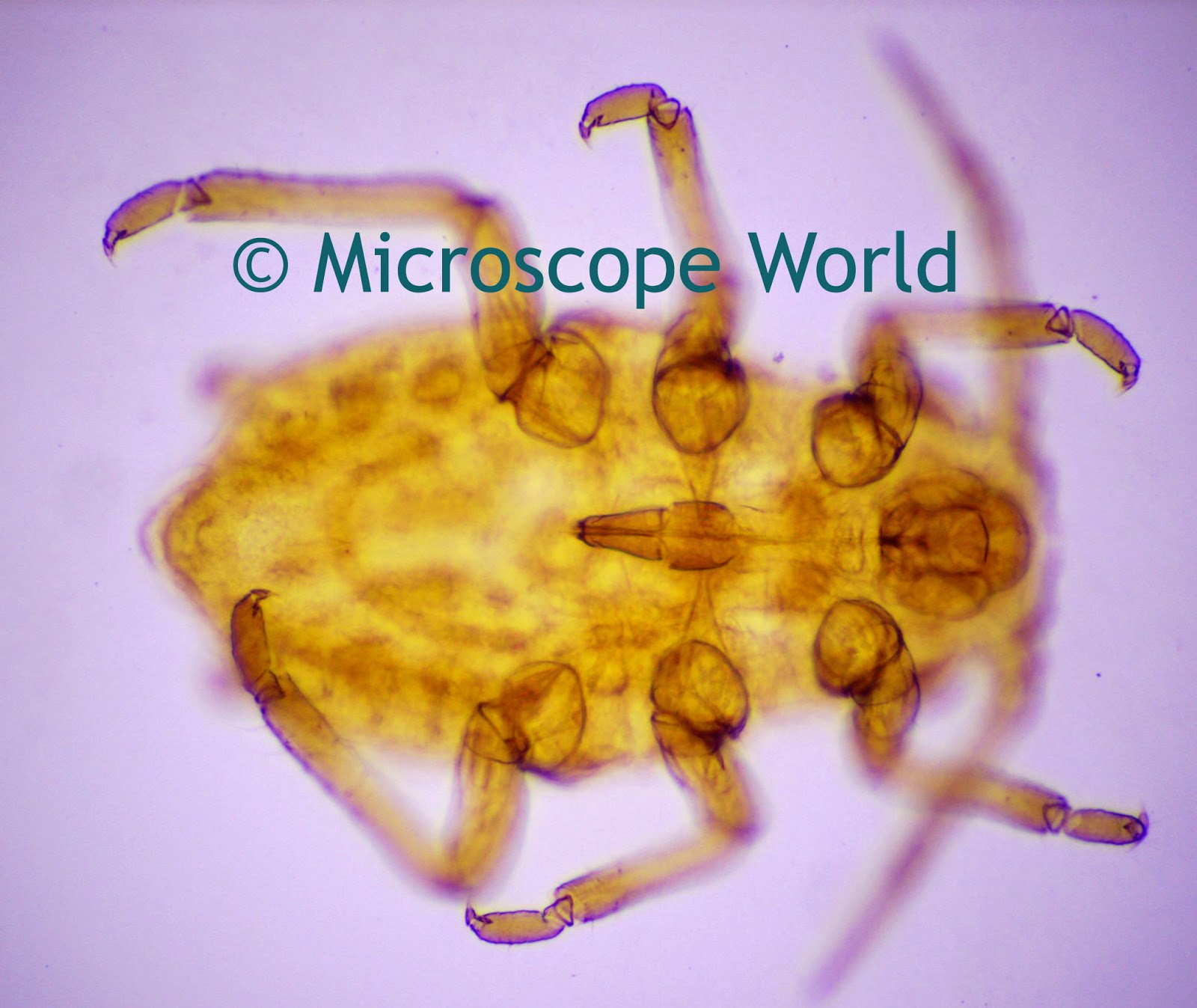
aphid under the microscope at 100x
Aphid captured at 100x magnification. (Example of a dry mount slide).


Wet Mount Microscope Slides

Wet mount slides are used to view liquids under the microscope. Preparation of a wet mount slide includes a depression slide, a cover slip, and an eye dropper can be useful, but is not required. Wet mount slide samples might include pond water, cheek cells, blood or sperm samples.

When preparing the wet mount slide, place a small drop of the sample between the depression slide and the cover slip. Use a paper towel when pressing the cover slip onto the slide, as some of your sample might run out over the edges. If you want to create a permanent slide, you can line the edges of your cover slip with clear nail polish. This will allow you to permanently affix it to the slide, locking the moisture into the chamber you create between the depression slide and the cover slip. If you make your slide permanent be sure to seal the entire edge of your cover slip so the sample does not evaporate over time.

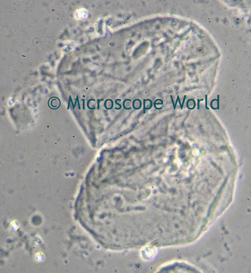
cheek cells under phase contrast microscope
Cheek cells captured under a phase contrast microscope at 400x. (Example of a wet mount slide).

If your sample requires staining you can learn about the different types of staining and how to stain your microscopy specimens here.
Special Microscope Objective Lens Inscriptions

$
0
0
Microscope objective lenses sometimes have special uses. Below you will find information on some of the inscriptions that you may find on your microscope objective lenses.

Microscope objective lenses (image)
  • NIC - this objective lens is preferred for use with Nomarski microscopy.
  • DIC - this microscope objective is used for differential interference contrast.
  • UV - Ordinary glass is relatively opaque to ultraviolet light wave lengths below 400 nanometers. UV objectives contain specially formulated glass elements and coatings in order to transmit a relatively high percentage of light of such wavelengths. These particular microscope objectives are useful for near ultraviolet excitation in reflected light fluorescence work. 
  • PL or NH inscription on a phase contrast objective - PL stands for positive low, a phase contrast example in which the specimen appears darker than the background of the field of view. NH is less commonly used and it stands for negative high. Negative high is a type of phase contrast where the specimen appears lighter than its background.
  • POL or SF - the microscope objective is strain-free and is required for high quality polarized light microscopy.

Printed Packaging Under the Microscope

$
0
0
Printed packaging is often placed under a microscope in order to view ink printing quality. Microscope World was contacted by a packaging printing company to help them configure a microscope system to analyze their printed inks.
macro zoom lens microscope system
Macro zoom Lens

A macro zoom lens microscope system was used to analyze the printed ink on the packaging shown below.
Packaging placed under the zoom lens.
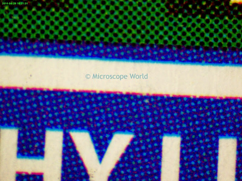
Ink under the microscope
Ink under the microscope.
The image above was captured from the packaging shown above. It is the text between the two words "Healthy Life". For more information on macro zoom lens configurations and custom solutions contact Microscope World.

Kids Microscope Ideas

$
0
0
Dissecting microscopes are great learning tools for younger kids, as just about anything found in the yard or around the house can be placed under the microscope for observation.

Here are some ideas of samples to look at under the microscope.
  • Pet hair versus human hair
  • Small insects - spiders, ants, gnats, beetles, etc.
  • Newsprint of package printing
  • Dollar bill
  • Flowers
  • Blades of grass
  • Pollen from a flower
  • Dirt and grease
  • Sugar and salt
  • Fabric and thread
  • Shoelaces
  • Coins and stamps
Fabric under the microscope
Fabric captured under the stereo microscope.
What do you like to look at under the microscope? Share images or ideas with us on the Microscope World Facebook page, we'd love to hear from you.

Metal Coating Inspection under Microscope

$
0
0
Microscope World recently had a customer who needed to view the consistency of coating on metal parts. Using the macro zoom lens microscope system, they were able to view and capture images at 15x and 160x magnification in order to inspect the layering (and pealing) of the metal coating.

metal under microscope
Metal piece for microscope inspection.
metal captured at 15x
Captured under the microscope at 15x magnification.
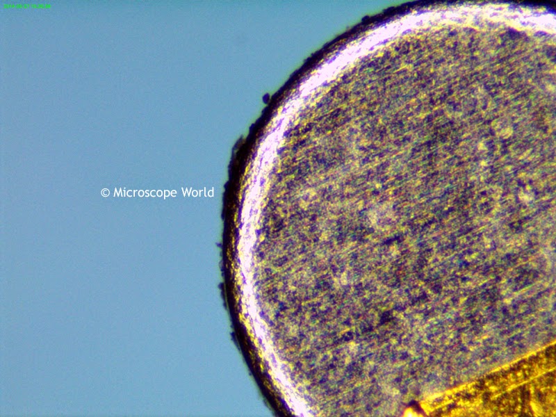
Microscope - metal at 160x magnification
Tip of a prong shown above captured at 160x magnification under the microscope.
All images were captured using a macro zoom lens, LED ring light and a 5 megapixel microscope camera. For custom macro zoom lens solutions please contact Microscope World.

Follow Microscope World on Google+

Searching for Metal Defects Under Microscope

$
0
0
Viewing metal under the microscope at higher magnification requires either a metallurgical microscope or a macro zoom lens microscope system. A metallurgical microscope is quite a bit more expensive than a macro zoom lens system.

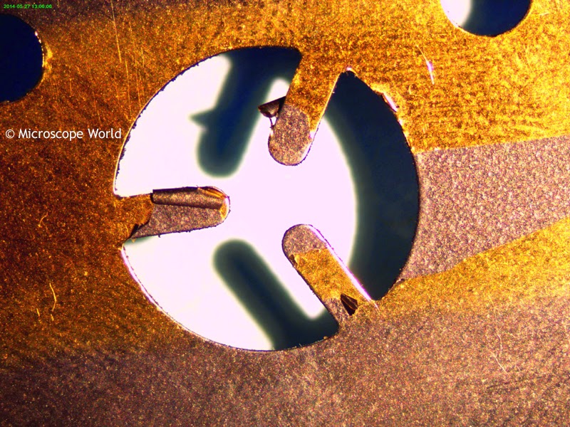
This metal plate needed to be inspected closely for defects. The area inspected under a macro zoom lens microscope was where the "Microscope World" text is located on the image shown above.

metal at 15x magnification
Metal captured under the microscope at 15x magnification.
Metal image at 160x under microscope
Metal captured at 160x magnification under microscope.
The higher magnification of the zoom lens was required in order to view defects in the metal plate.

Hydra Under the Microscope

$
0
0
Hydra is a genus of small, simple fresh-water animals, typically found in fresh-water ponds, lakes and streams in moderate and tropical regions. Hydra are usually a few millimeters long and are best seen under a biological microscope.

Hydra at 100x under microscope
Hydra captured at 100x magnification.
Each tentacle of a hydra is layered with highly specialized stinging cells called cnidocytes. The Cnidocytes contain specialized structures called nematocysts that, when contacted with prey, can fire neurotoxins paralyzing the prey.

hydra tentacles at 100x under microscope
Hydra tentacles captured at 100x under the microscope.
Hydra do not have a recognizable brain or muscles. The nervous system of Hydra is a nerve net.

hydra tentacles under the microscope (image)
Hydra tentacles captured at 400x magnification under the microscope.
Hydra extend their body to maximum length when feeding and slowly extend their tentacles. Upon contact with prey, the nematocysts on the tentacle fire into the prey while the tentacles coil around the prey.

Hydra 400x under microscope
Hydra tentacles connected to body of hydra, 400x under the microscope.
All hydra images were captured using the Richter Optica U2 biological microscope, with a 5mp microscope camera.

hydra tentacles under the microscope 100x
Hydra tentacles captured at 100x magnification.
Hydra captured at 400x under the microscope
Hydra body captured at 400x magnification under the microscope.
The hydra prepared slide can be purchased as part of the Zoology: Small Organisms prepared slide kit.

Science Project: Mold under the Microscope

$
0
0
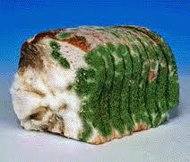
Mold is a fungus that grows in the form of multi-cellular filaments called hyphae. The best way to examine mold under a microscope is to grow your own in a controlled environment.  Soft bread without preservatives in it is perfect for this experiment. Leave the bread in a warm area, such as on the counter for a day, then place it in a ziplock bag with a small amount of water. Place the bread in a sunny area.


Once your bread is sufficiently moldy, wear a pair of gloves and put either a drop of water or a drop of Methylene blue (a staining agent) on a depression slide. Using a toothpick or a disposable knife, scrape some mold off the bread and place a small amount on the droplet of water. Cover it with a cover slip.

Using a high power student microscope to view the mold, start with the lowest magnification of 40x and work your way up to 400x.

Draw some images of your mold. Does it have a thin branch like structure? When you are finished with your project dispose of the moldy bread and be sure to wash your hands thoroughly.

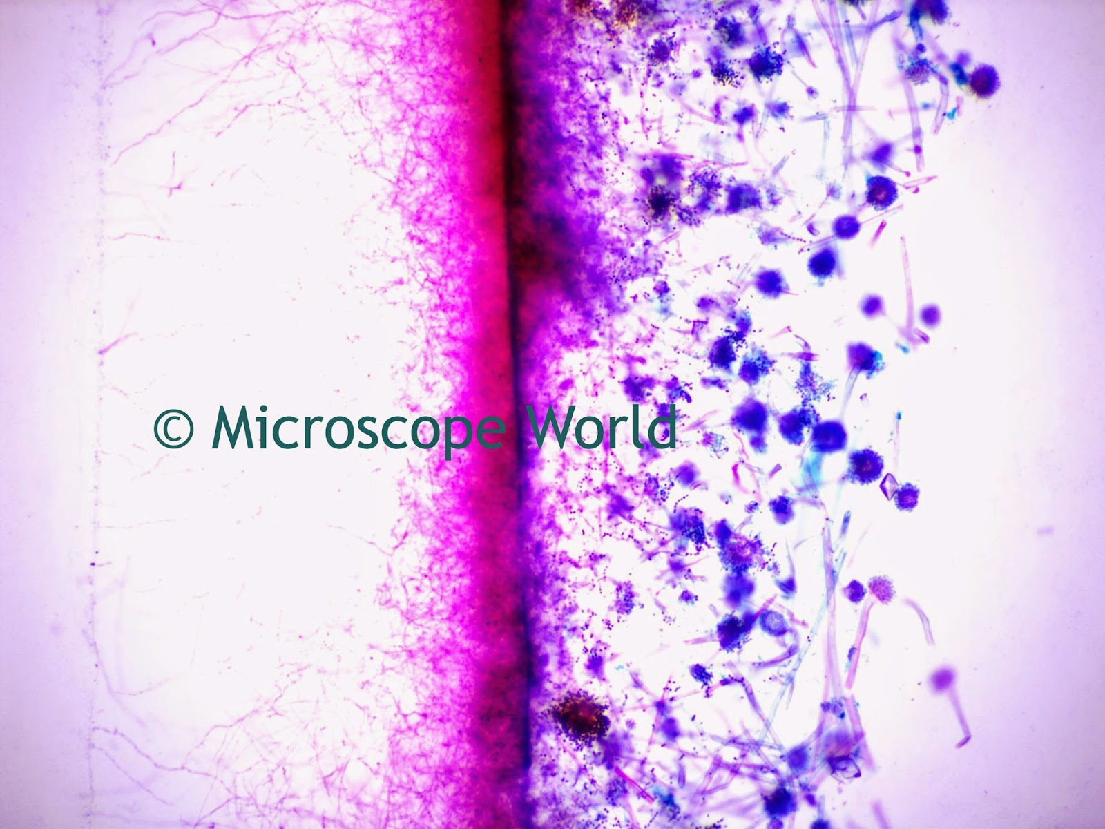
mold under microscope at 100x magnification
Mold under microscope, 100x

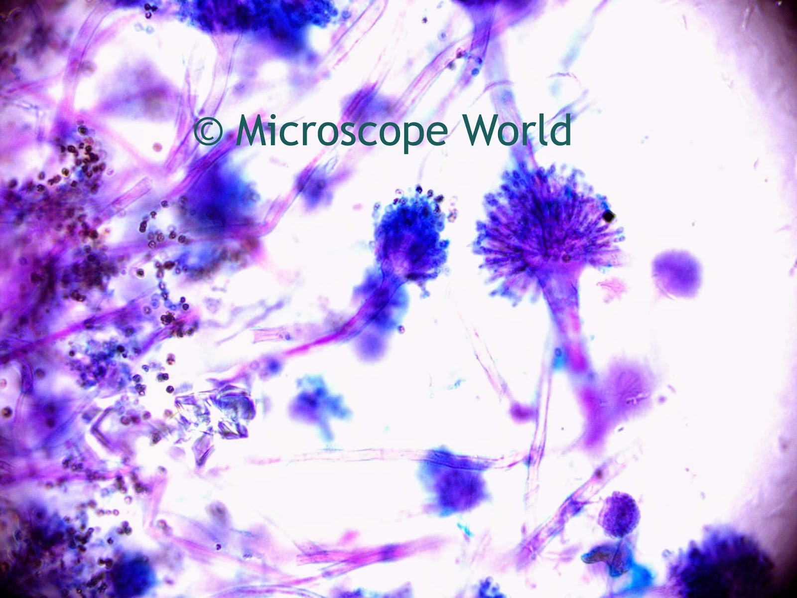
mold under the microscope at 400x
Mold under microscope, 400x


Closeout Special Microscope SALE!


Clam Worm Under the Microscope

$
0
0
Clam worms (Alitta Succinea) are a species of marine annelid, also known as sandworms. Clam worms can be found in the Northwest Atlantic region, the Gulf of Maine and South Africa. A clam worm can grow up to 6 inches in length, but usually they are a bit smaller. An interesting feature of the clam worm is the fact that they have four eyes.

Photo: Hans Hillewaert (Belgian North Sea)
The clam worm is a freeswimming polychaete that scavenges on the bottom of shallow marine waters feeding on other worms and algae. The clam worm itself is a food source for crustaceans and bottom-feeder fish.

clam worm under microscope
Clam worm prepared slide, captured at 40x magnification.
clam worm under microscope
Clam worm captured under the microscope at 100x.
microscope clam worm image
Clam worm captured under microscope at 400x magnification.
All images were capturing using the U2 biological microscope and the DCM5.1 microscope camera with 5 mega pixels.

Grantia under the Microscope

Radiolaria under the Microscope

$
0
0
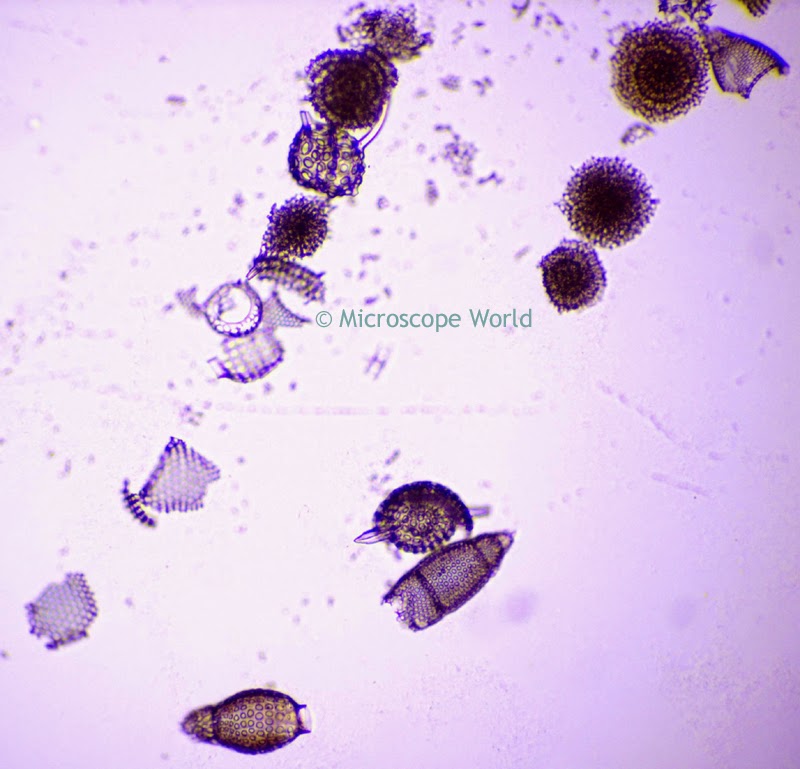
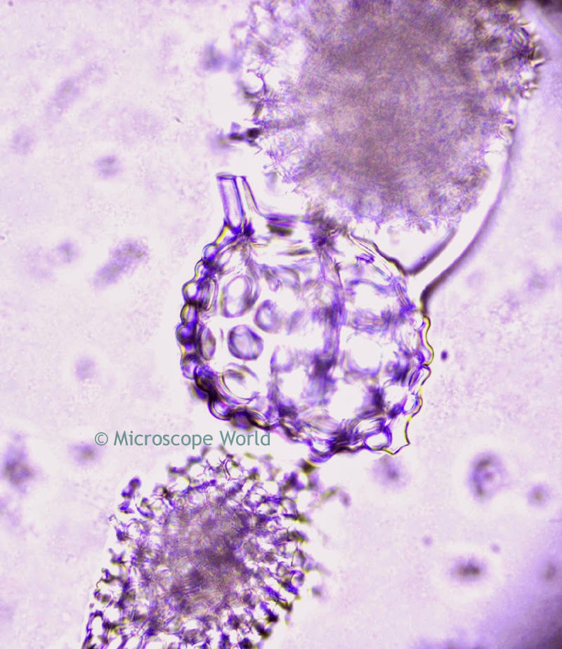
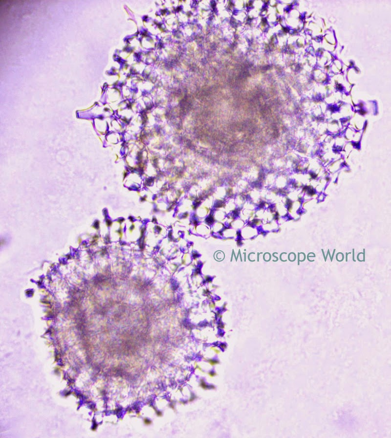
Radiolaria are protozoa that produce intricate mineral skeletons. These skeletons, found as zooplankton throughout the ocean, usually have a central capsule that divides the cell into inner and outer portions of endoplasm and ectoplasm. The cell nucleus is in the endoplasm, while the ectoplasm is filled with frothly vacuoles and lipid droplets. Radiolarians are incredibly buoyant due to their needle-like pseudopodia that are supported by bundles of microtubles.

Radiolaria under the microscope at 100x
Radiolaria captured at 100x magnification under a biological microscope.
These images of Radiolaria were captured using the Richter Optica U2 biological microscope with a 5 mega pixel microscope camera.

Radiolaria at 400x under microscope
Radiolaria, 400x magnification
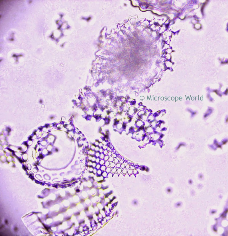
Ninety percent of radiolarian species are extinct. The skeletons of ancient radiolarians are used in geological dating, including for oil exploration and determination of ancient climates. Higher concentrations of dissolved carbon dioxide in sea water dissolves the Radiolaria's delicate structure, seen as fractured scattered pieces under a microscope.

Radiolaria under microscope 400x
Radiolaria, 400x magnification.
Radiolaria under microscope at 400x.
Radiolaria, 400x magnification.

How to Calculate Stereo Microscope Magnification

$
0
0
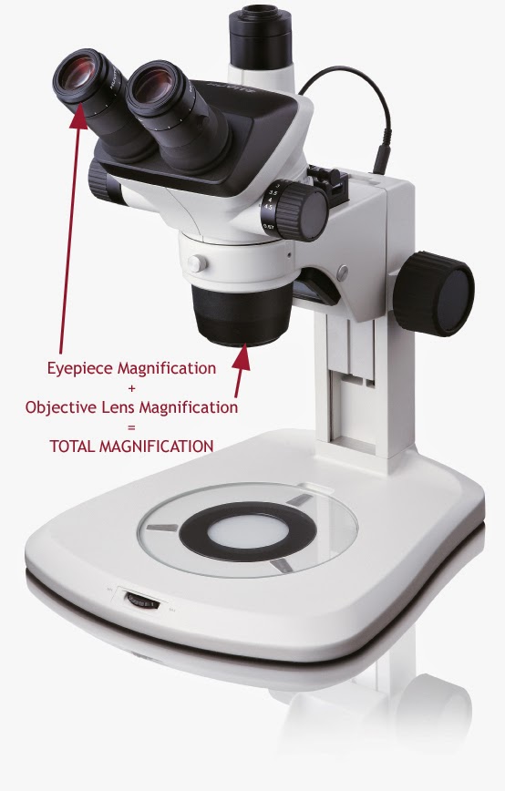
Stereo microscope magnification is a combination of the eyepiece magnification (most commonly 10x) and the objective lens magnification (typically anywhere between 0.7x - 5x). If you are using a stereo microscope with 10x eyepieces and the zoom knob is set to 4x, the total magnification formula would look like this:

10 x 4 = 40x magnification


stereo microscope magnification image
Eyepiece Magnification x Objective Lens Magnification = Stereo Microscope Magnification

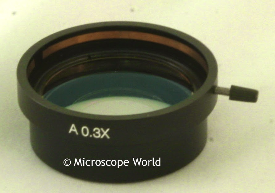
The other addition that can alter total magnification is if the stereo microscope has an auxiliary lens added to it. If this is the case, the auxiliary lens is also multiplied into the equation. Say you are using a stereo microscope with 10x eyepieces, the zoom knob is set to 5x and you also have a 0.3x auxiliary lens on the microscope. Total magnification would be determined with the following formula:

10 x 5 x 0.3 = 15x magnification

stereo microscope auxiliary lens image
Stereo Microscope Auxiliary Lens

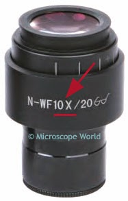
microscope eyepiece magnification
Microscope Eyepiece Magnification of 10x

Often the magnification of the microscope eyepiece will follow the letters "WF", depicting a Widefield eyepiece.


zoom microscope knob image
Stereo Zoom Microscope Knob for adjusting Zoom Magnification
The adjustment for the zoom objective lens value is found on the side of the microscope and the corresponding objective number is printed on the knob.

On a basic stereo microscope setup, to determine total magnification simply look at the magnification on the eyepiece and on the zoom knob. Stereo microscope auxiliary lenses are only usually used when the working distance needs to be adjusted or in some cases if magnification is being pushed quite high. For questions about stereo microscope magnification contact Microscope World.

Paramecium under the Microscope

$
0
0
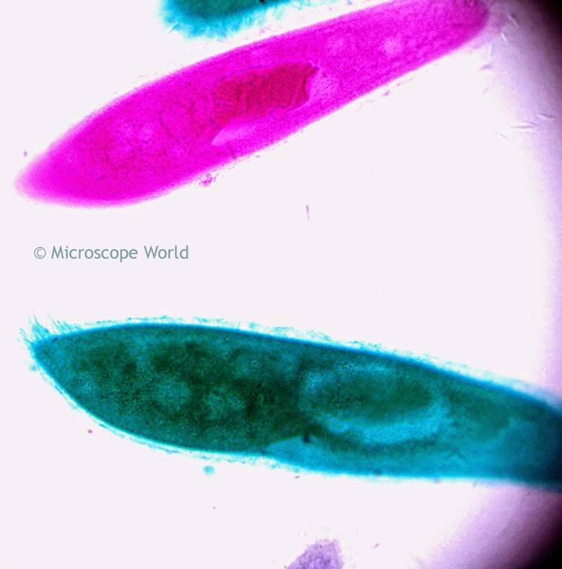
Paramecium is a genus of the single cell Ciliate Protozoa and are found in freshwater, marine areas, and often in stagnant ponds. Paramecium are unique to microscopy because they were one of the first ciliates to be seen by microscopists in the late 17th century.

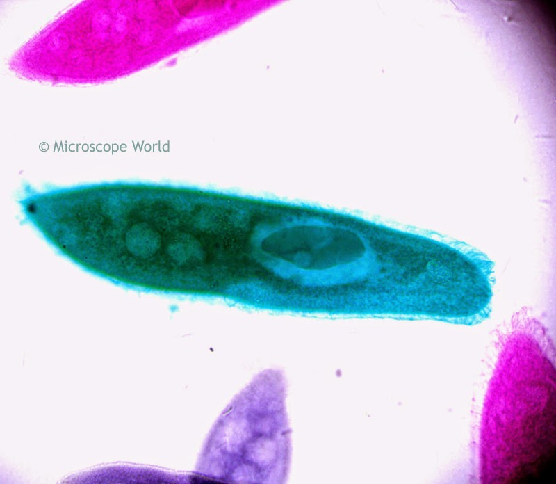
Paramecium under microscope at 400x
Paramecium captured under the microscope at 400x.

In 1718 the French math teacher and microscopist Louis Joblot published a description and illustration of a microscopic "Poisson" (translated fish) that he discovered in an infusion of oak bark in water. It was a Paramecium. He called it "Chausson" (translated slipper) and the term slipper animalcule remained in use as an everyday description for Paramecium throughout the 18th and 19th centuries.

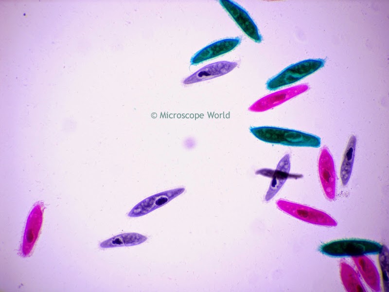
Microcope image of paramecium at 100x
Paramecium captured at 100x under the microscope.
The name "Paramecium" was constructed from the Greek paramekes and means "oblong".

Microscopic image of paramecium at 400x
Paramecium, 400x
All Paramecium images were captured using the U1 trinocular biological microscope and a microscope digital camera.
Viewing all 422 articles
Browse latest View live