Quantcast
Channel: Microscope World Blog
Viewing all 422 articles
Browse latest View live

Arcella Vulgaris under the Microscope

$
0
0
Arcella Vulgaris is a Testate Amoeba that has loose pseudopodia protruding out of the pseudochitin shell. This video shows the Arcella Vulgaris producing Carbon Dioxide vesicles to create buoyancy.  The test (or shell on sea urchins and microorganisms) is for protection and secreted by the animal itself. Testate amoeba are amoeba that have an umbrella shaped hard covering.


Arcella Vulgaris lives among water weeds and feeds on bacteria. The video also shows another variety of Arcella sp. which is transparent and the Amoeba inside can clearly be seen.

In the video captured by Ravindra Bakare, Associate Professor of Zoology at Kisan Veer Mahavidyalaya in Wai Dist Satara, Maharashtra, India, stagnant pond water was collected near the University and placed under a biological microscope. Bakare works for the fresh water protozoan diversity around Wai and captured this Arcella Vulgaris video under the microscope by using a 2 megapixel eyepiece camera at 45x magnification. Microscope World is grateful for Ravindra Bakare's hard work in capturing this video and for sharing it with microscopy enthusiasts!
Comparator Reticles

$
0
0
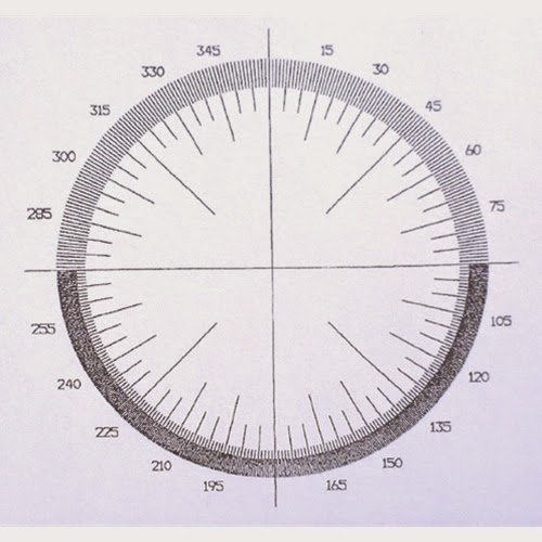
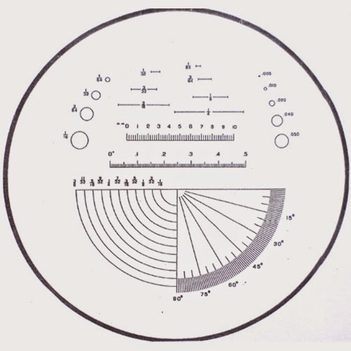
Microscope comparator reticles for use in a biological or stereo microscope are available in full 360°, half comparators (180°), and comparators for specific objectives (1x, 2x, 3x). The microscope comparator reticles are available in diameters of 16mm - 28mm.

Microscope comparator reticles, available in 360 or 180 degrees.
360 Degree Comparator Microscope Reticle
The standard comparator reticle is available in 26mm - 28mm diameters and contains rulers, circles of varying diameters and comparator protractors with different angles.

Microscope standard comparator reticle for measuring.
Standard Comparator Reticle

Best Cordless Field Microscopes

$
0
0
We reviewed many cordless microscopes and below have listed the best cordless field microscope options.

Best Cordless Kids Microscope

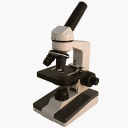
Elementary Kids microscope Richter Optica F1.
The Richter Optica F1 kids microscope is hands down the best option for a cordless kids microscope. This microscope uses a cool LED light - this is important in order to keep live specimens from getting killed by the heat of the microscope light! The microscope has coarse AND fine focusing - very important in order to view a clear image at 400x. Glass optics = clear images. Magnification of 40x, 100x, 400x - high enough to view bacteria, blood cells and Protozoans in pond water. Lightweight at only 3 lbs. and easy to carry. The Richter Optica F1 sells for $99.


Best Stereo Zoom Dissecting Cordless Microscope

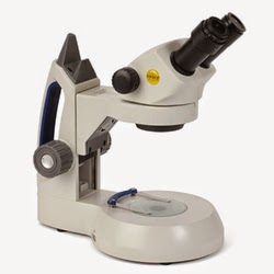
Swift Optical SM105 stereo dissecting microscope is cordless.The Swift Optical SM105 stereo zoom microscope provides continuous magnification of 10x - 30x, which is
perfect for viewing flowers, rocks, insects and any other item that might be found out in the field and held in the palm of your hand. Cool LED light won't heat up. Top light and bottom light can be operated independently of each other. Carrying handle makes this dissecting microscope easy to transport. The Swift SM105 stereo microscope sells for $489.


Best Plan Achromat Compound Binocular Cordless Microscope

Richter Optica UX-1 Binocular Compound Microscope with Plan Achromat Objectives
The Richter Optica UX-1 is a great advanced high school microscope. With Plan Achromat objective lenses the full field of view is completely flat, crisp and clear. Binocular viewing makes it easier to view specimens through the microscope and the mechanical stage makes maneuvering these samples even easier. Magnification of 40x, 100x, 400x and 1000x provides standard lab magnifications for viewing everything from bacteria and cells to pond life creatures. Without any cords to worry about, the UX-1 is also quite popular among veterinarians. The UX-1 compound microscope sells for $699.


Best Tablet LCD Cordless Microscope

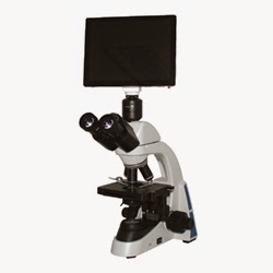
Cordless Tablet LCD microscope with WiFi.The UX1-LCD tablet microscope has all the great features of the UX-1 shown above, but it also
includes a 10" LCD Tablet camera. The camera has a 5 mega pixel camera and an SD card slot, as well as HDMI output. The Android tablet provides a touch screen and measurements can be made as well. The UX1-LCD tablet microscope sells for $1989.

Earth Day under the Microscope

$
0
0
Today in honor of Earth Day, we have decided to share some of our favorite "earth" microscopy images. Enjoy!

Tremolite under the polarizing microscope.
Tremolite under a polarizing microscope.
Tremolite is a mineral that forms from metamorphism of sediments rich in quarts and dolomite.

Monocot and Dicot slide under the microscope at 40x magnification.
Monocot and Dicot (flower) captured under a biological microscope at 40x.

Moss under a biological microscope at 400x.
Moss captured under a biological microscope at 400x.

Zebrafish under a stereo fluorescence microscope, day 3.
Zebrafish (day 3) under a stereo fluorescence microscope.

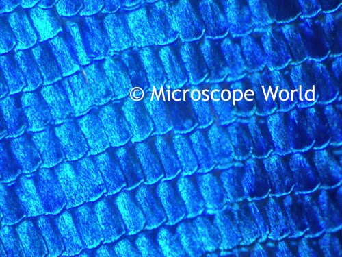
Butterfly wing under the microscope at 100x.
Butterfly wing captured at 100x magnification under a biological microscope.
Happy Earth Day!

Geology Microscope Images

$
0
0
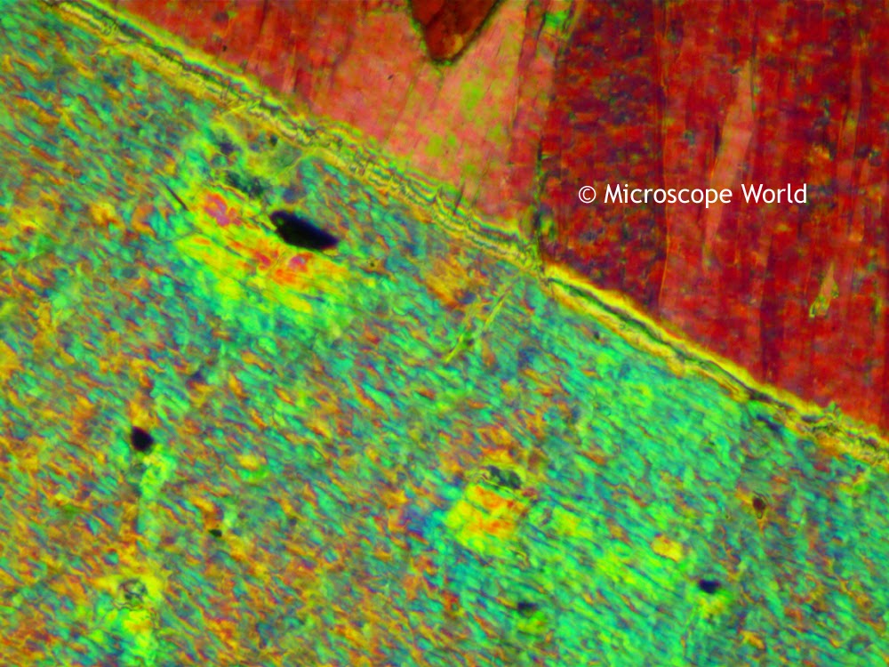
The thin sections of rock shown below were captured using the Fein Optic RB40 polarizing microscope. All images on this page were captured using the DCM3.1 microscope camera (3 megapixels).

Geologist microscope image using the 1/4 wave plate.
Geology thin section using the 1/4 wave plate under the R40POL polarizing microscope.

Polarizing microscope image using the full wave plate.
Geology thin section using the full wave plate and full polarization under the R40POL polarizing microscope.

Polarizing microscope image of a thin section using the full wave plate.
Geology thin section using the full wave plate and full polarization under the R40POL polarizing microscope.

Polarizing microscope image of a thin section of rock using the full wave plate.
Geology thin section using the full wave plate under the R40POL polarizing microscope.

Geology microscope image of a thin section of rock using a polarizing microscope.
Geology thin section using polarization with the R40POL polarizing microscope.

Polarizing microscope image of a thin section of rock using polarization technique.
Geology thin section using polarization with the R40POL polarizing microscope.

Diadema Larvae Under the Microscope

$
0
0
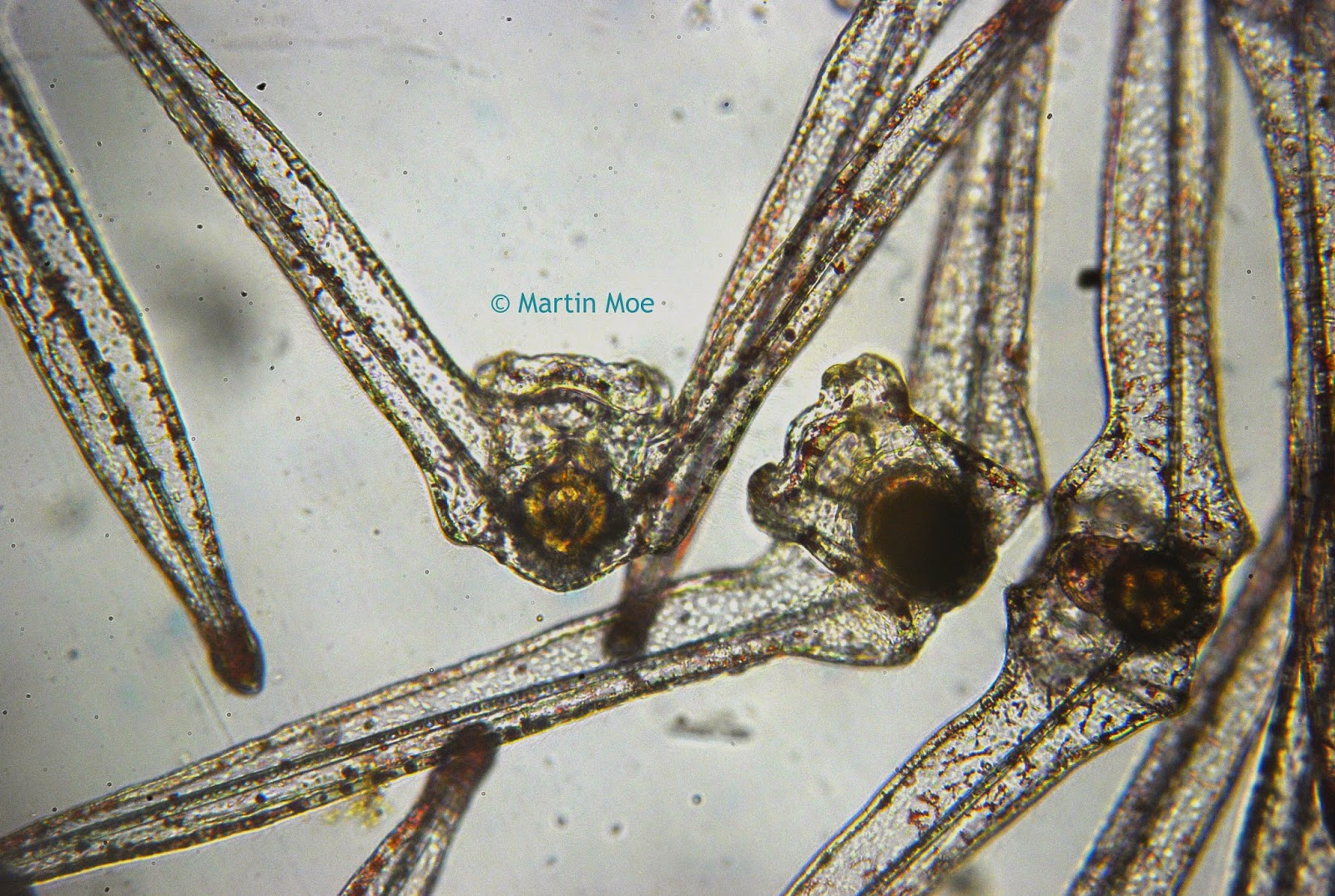
The microscope image below is of a 13-day-old Diadema larvae. Diadema larvae are an interesting sea urchin larvae. Two of the spines that are typical of other sea urchin larvae actually grow into centimeter long "wings" and they glide through the ocean currents for possibly months before finding a place to settle and metamorphose into tiny juvenile Diadema sea urchins.

Diadema Larvae captured under the microscope, Day 13.
Diadema Larvae, Day 13. Image courtesy: Martin Moe
A big thanks to Martin Moe for capturing this image (with the Meiji MT4000 series biological microscope) and sharing it with Microscope World!

Protozoans under the Microscope

$
0
0
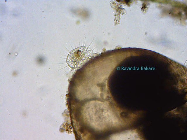
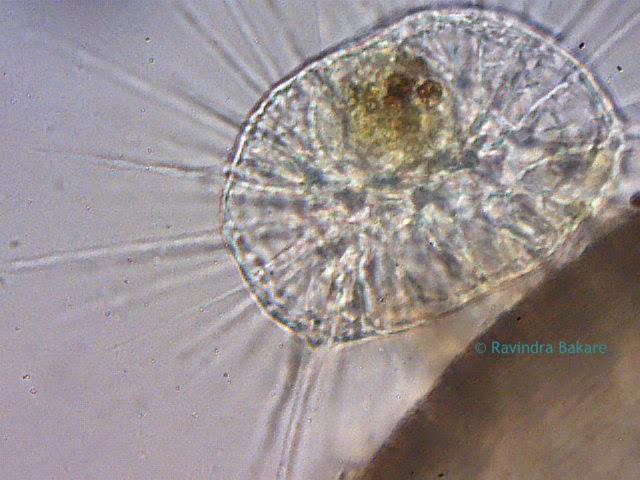
The protozoan shown below is Actinosphaerium sp. This protozoan adjusted itself on the surface of a shell of a freshwater gastropod. The Actinosphaerium sp. is a kind of Amoeba with a needle like pseudopodia used for catching food.

Protozoan Actinosphaerium sp. under the microscope at 10x.
Actinosphaerium sp. at 10x magnification. Image courtesy: Ravindra Bakare.

Actinosphaerium sp. protozoan and amoeba under the microscope.
Actinosphaerium sp. at 45x magnification. Image courtesy: Ravindra Bakare.

Images were captured by Ravindra Bakare, Associate Professor of Zoology at Kisan Veer Mahavidyalaya in Wai Dist Satara, Maharashtra, India. Stagnant pond water was collected near the University and placed under a biological microscope. Bakare works for the fresh water protozoan diversity around Wai and captured this Actinosphaerium sp. under the microscope by using a 2 megapixel eyepiece camera at 10x and 45x magnification. Microscope World is grateful for Ravindra Bakare's work in capturing these images and for sharing them with microscopy enthusiasts!
Microscope Mechanical Stages

$
0
0

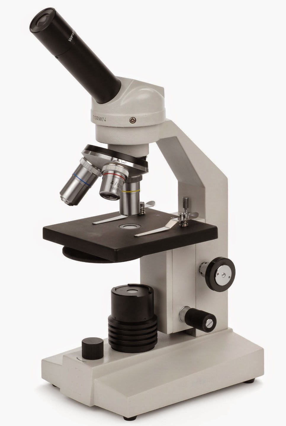
HS-1M High School Microscope

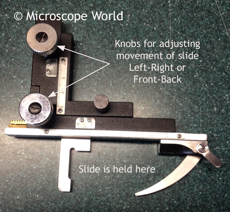
Microscopes often have a built-in mechanical stage, while others have the option to add a mechanical stage. A microscope mechanical stage allows the user to maneuver the samples under the microscope left and right or forward and backward simply by turning a small knob. A microscope mechanical stage gives the user much more control when viewing the sample and makes it easier to keep that sample inside the microscope field of view.

The mechanical stage shown below replaces the stage clips on the HS-1M high school microscope shown at left. By adding the mechanical stage it becomes easier to maneuver slides and keep samples in the field of view.

Microscope mechanical stage examples.
Attachable microscope mechanical stage.

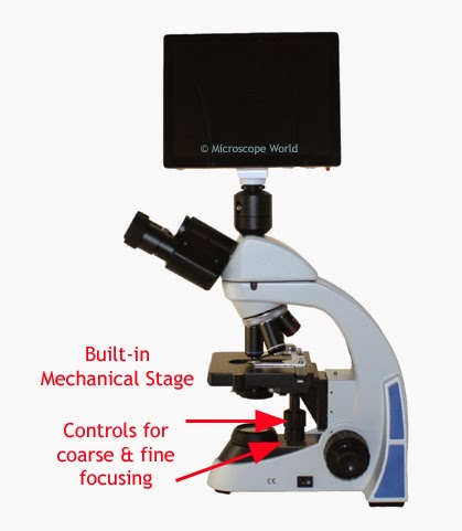
The UX1-LCD microscope below has a built-in mechanical stage. Notice the drop-down controls on the side of the microscope. One of these moves the stage left-right and the other moves the stage forward-backward.
Built-in microscope mechanical stage.
Built-In Microscope Mechanical Stage


The stereo microscope mechanical stage shown below is made for a stereo microscope with transmitted light.

Stereo microscope mechanical stage.
If you have questions about adding a mechanical stage to your microscope, please contact us.

Toolmaker's Microscopes

$
0
0
Toolmaker's microscopes are used for inspection and measurement of machined parts. The toolmaker's microscope is typically used in the quality control process when precise measurements are required.

Toolmaker's microscope with digimatic micrometer heads.
Toolmaker's microscope with digimatic measuring heads.

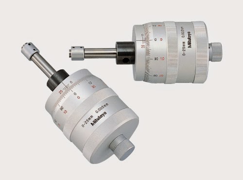
Toolmaker's microscopes are available with manual measuring micrometer heads, as shown below, or with digimatic micrometer heads. The manual micrometer heads are less expensive and provide measurements in either mm or inches. Measurements are made by positioning the sample in the center of the cross-line reticle in the microscope, then turning the micrometer (which controls the stage) until the edge of the part lines up with the crossline. The micrometer head measures the distance the stage moved.

Toolmaker's microscope manual micrometer measuring heads.
Manual micrometer measuring heads found on toolmaker's microscope.

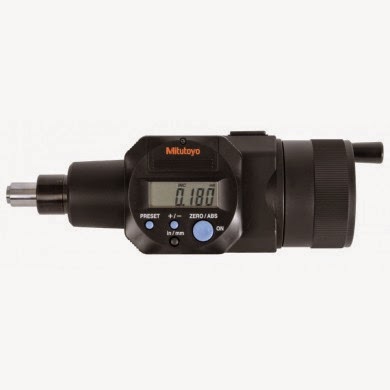
Digimatic micrometer measuring head for toolmaker's or measuring microscopes.
Digimatic micrometer measuring head found on toolmaker's microscope.

The digimatic micrometer measuring head fits on the stage of the toolmaker's microscope and performs the same functions as the manual micrometer head but with a digital readout. The digimatic micrometer measuring head can easily switch back and forth between measurements in mm and inches as well.

Microscope Troubleshooting

$
0
0
Getting a microscope to work can be frustrating. Below are a few pointers on how to troubleshoot some areas that might be causing you microscopy grief.

The Microscope Won't Focus

Microscope condenser image
Microscope Condenser
If you're using a biological compound microscope and can't seem to get the lenses into focus (in
particular the 40x and 100x objectives), check these areas:
  • The height of your condenser may be set too high or too low (this can also affect resolution).
  • Make sure that your objective lenses are screwed all the way into the body of the microscope.
  • On high school microscopes, if someone adjusts the rack stop, the microscope will not focus. The microscope rack stop is in place in order to prevent the lenses from slamming into the microscope stage and breaking. However, if it is out of place it will prevent the microscope from focusing properly.

Rack stop on high school microscope.

Microscope Rack Stop


If you are using a stereo microscope and can't get your image into focus, the body of the microscope is either too far away or too close to your object. If you know the working distance of the microscope, this is the distance that is required between the lens of the microscope and the top of your object in order for your sample to be in focus.

Microscope Working distance illustration
Microscope Working Distance

The Camera Won't Show an Image

If you can't see an image in the microscope camera, or the computer your camera is hooked up to only shows a black screen, make sure you pulled out the beam splitter on the microscope. The beam splitter sends light from the microscope eyetubes and directs it up to the camera.

Microscope beam splitter sends light to the microscope camera.
Microscope Beam Splitter

The Microscope Light Won't Work

  • Most microscopes have an on/off switch as well as a rheostat control. Make sure the rheostat is turned up. Many times the microscope is turned on, but the rheostat is off and won't allow light to pass through.
  • Is the field iris opened up?
  • Is your fuse burned out?
  • Is your microscope bulb burned out?
  • If you're using a cordless microscope, has it been charged?

The Stereo Zoom Microscope Won't Change Magnification

Stereo zoom microscopes adjust the magnification when the zoom knob is turned as gears inside the microscope turn. Over time and extended use these gears can sometimes become stripped. Below is an image of a stereo zoom microscope where the zoom magnification gears became stripped and the microscope would no longer change magnification. This type of problem requires professional technical repair or replacement. Contact your microscope manufacturer.

Internal stripped gears on a stereo zoom microscope.

There are Dirty Specks in the Microscope Field of View

Dirt on objectives, eyepieces, or in the internal parts of the microscope can result in microscopy images that are less than ideal. In order to determine which part of your microscope is dirty, and ultimately which parts you need to clean, follow these guidelines.
  • Look through the microscope while turning the eyepieces. If the spec of dirt moves, clean your eyepieces. (View cleaning tips here).
  • While looking through your microscope, move the slide on the stage. If the speck of debris moves, your slide should be cleaned, or your sample replaced.
  • If you have adjusted both the slide and the eyepieces and the speck of dirt does not move, clean your objective lens. Especially if you are using a 100x oil immersion objective, many times oil and dirt will build up on the objective. Microscope cleaning supplies can be found here.
When your microscope is not in use cover it with a dust cover. Store the microscope in a cool, dry place.

Microscope image captured with clean objective lenses
Clean Lens Microscope Image
Dirty microscope lens image
Dirty Lens Microscope Image
If you are having trouble with your microscope and can't figure out why it is not working, contact Microscope World.

Kids Science Project: Boiling Water

$
0
0
Ever wonder how much bacteria is killed by boiling water? This is great science project that will help you answer this question!

Science Project Supplies

Find a local stream or pond and fill your container with water. You don't need to gather mucky, dirty water - clear water is fine. Place a drop of the water on your depression slide and look at it under the microscope - specifically at 100x and 400x magnification. Can you locate some bacteria? The image below shows some of what you might find in your collected water.

Bacteria under the microscope.
Next, fill a pot with your pond water and boil the water for a minimum of 30 minutes. Once the water has cooled, place a drop of your boiled water under the microscope. Have the bacteria disappeared? Would you feel safe drinking this water? What remains under the microscope after the water was boiled?

If you are presenting your science project to your class, show images or drawings of what was under the microscope both before and after you boiled the water. Explain what caused the water contents to change.

Richter Optica S6.7 Stereo Zoom Microscopes

$
0
0
Richter Optica has updated the S6.6 stereo zoom microscope to the S6.7 stereo zoom microscope. This new stereo microscope release includes many of the same features from the S6.6 stereo microscope with several added benefits. New features of this stereo zoom microscope include:
  • Dual focusing widefield eyepieces (available in 10x, 15x and 20x)
  • 10x & 15x eyepieces accept 24mm diameter reticle
  • Zoom range of 0.67x - 4.5x
  • Bright dual LED top and bottom illumination with individual rheostat controls
  • Optional 0.5x, 1.5x and 2x auxiliary lenses
  • Optional polarizing set for the stereo microscope
  • Optional darkfield kit for the stereo microscope
  • Binocular and trinocular microscopes available
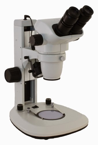
Richter Optica S6.7 Stereo Zoom Microscope with 6.7x - 45x magnification and dual LED lights.
Richter Optica S6.7 Stereo Zoom Microscope
 If you have questions regarding the Richter Optica S6.7 stereo zoom microscope contact us.

How Does a Light Microscope Work?

$
0
0
Ever wonder how a light microscope (also known as a compound microscope or biological microscope) works? This article explains in detail how a light microscope works.

A compound light microscope gathers light from a small area (where your specimen is on the stage) and sends this light up through the objective lens. The objective lens magnifies the sample, as do the eyepieces you are looking through.

Image explaining how a light microscope works.
Light Microscope Features and Functions

In order to focus the image, the coarse focusing is used first in order to put the sample in the correct location to obtain a clear image. On light microscopes, moving the focusing knob will either move the stage up and down, or move the head of the microscope up and down. On the U2 biological microscope shown above, moving the coarse focus adjusts the height of the stage. On most high school microscopes, the focusing mechanisms moves the height of the head of the microscope. Once the coarse focus knob has been used to put the sample in the correct location, next the fine focus knob is used to fine-tune the focus and create a crisp and clear image.

Light microscopes use a condenser above the light source in order to focus and direct the light up through the specimen (in a small beam of light) and into the eyepieces. Both the rheostat on the light as well as the iris on the condenser are used in order to get this beam of light focused properly when viewing the sample.

The objective lenses can be rotated in order to change the magnification of the microscope. Most light microscopes utilize 10x eyepieces and this magnification is multiplied by the objective lens value to determine the microscope magnification. For example, when using the 4x objective lens (4x10=40x) the magnification is therefore 40x. If the objective lens is rotated to the 10x objective (10x10=100x) the total magnification becomes 100x.

Light microscopes provide magnification up to 1000x. Any light microscope that advertises magnification above 1000x will be providing empty magnification and the images will not be clear or in focus.

Mitutoyo Objectives and Microscopes

$
0
0
Mitutoyo


Microscope World is proud to carry the entire Mitutoyo line including objective lenses, toolmaker microscopes, profile projectors, hardness testers, calipers, micrometers, etc. Mitutoyo microscopes and industrial metrology products have been manufactured to meet high quality standards for over 80 years. All products are manufactured in Japan.


Mitutoyo is known for their incredibly high quality (including high NA) objective lenses. All of the Mitutoyo objective lenses can be purchased online. Many of the Mitutoyo objectives are suitable for laser use.

The Mitutoyo products can be found online here. If you are unable to locate a specific Mitutoyo product, please contact Microscope World.

Remember

$
0
0
USA flag memorial day.


Today we pause to remember those who have served our country. Thank you!

Kids Science Project: Bottled Water versus Tap Water

$
0
0
Ever wonder if bottled water is cleaner than the tap water from your home? This is a great experiment that can help you find out!

Science Supplies Needed for Science Project:


Richter Optica F1 kids microscope
Kids Microscope F1
Prepare two different slides - one with your bottled water and one with the tap water and place each under the microscope. Can you identify any different particles in one versus the other? You will want to take a minimum of five samples from each the bottled water and the tap water to get a good sample size. Make sure you keep track of your results and draw or capture images of what you see in the water.

Bacteria can be viewed at 400x magnification, as well as most living organisms. Which water are you more comfortable drinking? Do you think there is a big difference between tap water and bottled water? You may also want to try this experiment in different locations - for example if you travel to a different state to visit friends or family you may want to bring some tap water home to test out your experiment and hypothesis again.


Birth Control Pill Under the Microscope

$
0
0
Ever wonder what the birth control pill looks like under the microscope? Maurice Mikkers started his career in laboratory research where he worked in the Netherlands for the National Institute for Public Health and the Environment. He eventually traded in his lab coat for a camera and is now working as a freelance photographer.

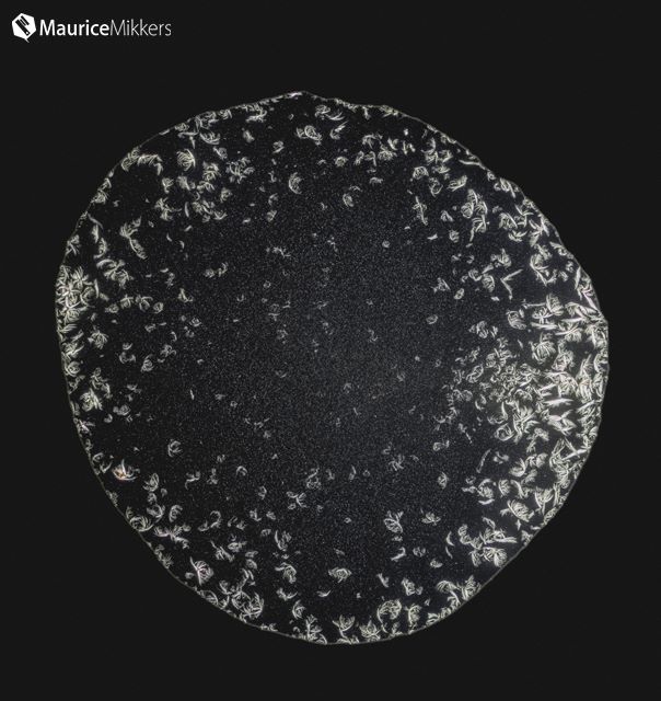
Mikkers prepared some microscope slides with the birth control Ethinylestradiol / levonorgestrel 0.03/015 pills. After 13 hours, his prepared slides were crystallized and he captured the photographs below using a Canon EOS 5D Mark III 22 megapixel camera mounted on a polarizing microscope.

Image of birth control pill under a polarizing microscope.
Birth Control Ethinylestradiol/levonorgestrel 0.03/0.15 under a polarizing microscope. Image Courtesy Maurice Mikkers.

Polarizing microscopy image of birth control pill captured by Maurice Mikkers.
Birth Control Ethinylestradiol/levonorgestrel 0.03/0.15 under a polarizing microscope. Image Courtesy Maurice Mikkers.


Thank you to Maurice for sharing his microscopy images with Microscope World. He has captured a number of different images of drugs and medicine under the microscope and you can view more images by Maurice Mikkers here.

Bacillus Subtilis under the Microscope

$
0
0
Bacillus Subtilis is also known as hay bacillus or grass bacillus. It is a gram-positive bacterium found in soil and the GI tract of ruminants and humans. Bacillus subtilis is rod-shaped and typically 4-10 microns long. This bacterium can form a tough, protective endospore that allows it to tolerate extreme environmental conditions.

Bacillus Subtilis captured at 40x under a biological microscope.
Bacillus Subtilis captured under the U2 biological microscope at 40x.

Bacillus subtilis is considered one of the best studied gram-positive bacterium and a model organism for studying bacterial chromosome replication and cell differentiation. It is one of the bacterial champions in secreted enzyme production and is used on an industrial scale by biotechnology companies.

Bacillus subtilis under the U2 biological microscope at 100x magnification.
Bacillus Subtilis captured under the U2 trinocular microscope at 100x.

Commonly found in the upper layers of the soil, bacillus subtilis can divide symmetrically to make two daughter cells (binary fission), or asymmetrically, producing a single endospore that can remain viable for decades and is resistant to unfavorable environmental conditions such as drought, salinity, extreme pH, radiation and solvents.

Microscopy image of bacillus subtilis at 100x magnification.
Bacillus Subtilis captured under the U2 biology microscope at 400x.
Bacillus subtilis is only known to cause disease in severely immuno-compromised patients and can conversly be used as a probiotic in healthy individuals. The bacteria rarely causes food poisoning.

Bacteria under the microscope captured at 400x using a plan fluor microscope objective lens.
Bacillus Subtilis captured using the Plan Fluor 40x Microscope Objective on the U2 microscope.

Measuring Calipers

$
0
0
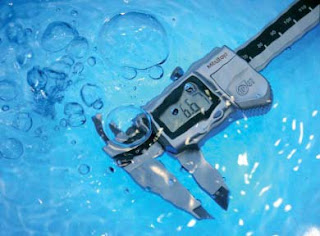
Microscope World now offers measuring calipers for measuring a variety of inside and outside diameters on tubes, pipes, metal fabrication and industrial manufacturing. The calipers are available in vernier as well as digital (mm and inches), and even waterproof and coolant proof versions.

Vernier caliper used for measuring outside diameter.
Vernier caliper measuring outside diameter.

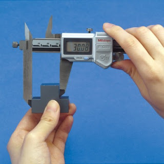
Vernier caliper used to measure inside diameters in industrial manufacturing.
Vernier caliper measuring inside diameter.

Measuring calipers for offset measurements in stepped sections.
Offset digital caliper.

The digital calipers have several options for downloading the data measurements directly to a computer either with a USB cable, or with a wireless signal.

Digimatic caliper for measuring and exporting data.
Digimatic caliper for exporting measurement data.


Calipers for measuring and exporting digital data are waterproof and coolant proof.
Waterproof and coolant proof digital caliper.

All measuring calipers can be found here and most categories contain options for both vernier or digital options. If you are looking for a specific type of caliper and can't locate it please contact Microscope World, as not all calipers available are on the website.

Petrographic Examination of Aggregates for Concrete Microscopes

$
0
0
Petrographic examination of aggregates for concrete, mortars, and building stones can help determine if the concrete cured correctly, of if there is any evidence of degradation. Concrete and aggregate testing provides construction managers valuable information during a project including concrete mix, air content, weight, and strengths of concrete.

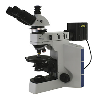
The Petrographic Examination of Aggregates for Concrete follows guidelines for petrographic microscopes that meet ASTM C295, ASTM C547M and ASTM C856 standards. Both concrete examination microscopes offered by Microscope World meet these standards.

View the petrographic examination of aggregates for concrete microscopes here.

ASTM C295, C457M C856 petrographic examination of aggregates for concrete microcope.
Petrographic Exam Aggregates Concrete Microscope
ASTM C295, ASTM C457M, ASTM C856

The Fein Optic R40PEAC polarizing petrographic microscope for examination of aggregates for concrete provides transmitted light along with high quality strain-free polarizing objectives for the most intensive aggregate and concrete examination work. This system meets standards for ASTM C295, ASTM C457M and ASTM C856.

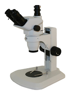
ASTM C295, C457M and C856 petrographic stereo microscope for aggregates / concrete.
Petrographic Stereo Microscope
The Fein Optic FZ6PEAC polarizing stereo microscope provides magnification of approximately 6x-150x. This microscope system meets standards for ASTM C295, ASTM C457M and ASTM C856.
Viewing all 422 articles
Browse latest View live