Quantcast
Channel: Microscope World Blog
Viewing all 422 articles
Browse latest View live

Stereo Boom Stand Microscopes

$
0
0
Stereo microscope are often used to inspect large parts. When using a standard stereo microscope, often times the large pieces will not fit under the microscope. This is when a boom stand becomes helpful.

Stereo microscope on ball bearing boom stand.
There are three types of microscope boom stands.
  1. Standard boom stands have a single horizontal and vertical pole. 
  2. Ball bearing boom stands slide on the horizontal axis, making it easier to move the stereo microscope head back and forth when viewing objects.
  3. Articulated arm stands have several joints that can be locked into place.
Stereo microscope on articulated arm stand.

Cork under the Microscope

$
0
0
Microscope World recently used the HSZ6-TBL stereo zoom microscope to view a cork from a wine bottle.

Cork is a buoyant material that is harvested for commercial use primarily from the Cork Oak, which is found in southwest Europe and northwest Africa. Cork is impermeable, buoyant, and fire resistant and is therefore used in a variety of products, the most common of which are wine stoppers. The montado landscape of Portugal produces approximately 50% of cork harvested annually worldwide.

100x Magnification obtained with higher magnification c-mount adapter.
100x Magnification. Captured with DCM2.1 microscope camera.
100x Magnification.
You will notice in the above images of cork that some of the crevices are not fully in focus - this happens because the higher the magnification on the microscope, the smaller the depth of field becomes. So if you are not working with a substance that is completely flat, you will have to refocus in order to see hills and valleys in the specimen.

England Finder Slide

$
0
0
The England Finder is a glass slide marked in vacuum deposited chromium over the top surface in such a way that a reference position can be deduced by direct reading, the relationship between the reference pattern and the locating edges being the same in all finders. The object of the Finder is to give the microscope user an easy method of recording the position of a particular field of interest in a specimen mounted on a slide, so that the same position can be re-located using any other England Finder on any light microscope.

England Finder Illustration

The England finder slide consists of a glass slide marked with a square grid at 1mm intervals. Each square contains a center ring bearing reference letter and number, the remainder of the square being subdivided into four segments numbered 1-4. Reference numbers run horizontally 1-75 and letters vertically A-Z (omitting I). The main locating edge is the bottom of the slide which is used in conjunction with either the left or right vertical edge of the slide, according to the fixed stop of the stage of the microscope, all three locating edges being marked with arrow heads. The label on the finder should always appear visually at the bottom left corner when through most microscopes the reference image will appear correct. You can purchase the England Finder Slide here.

Stereo Fluorescence Microscopes

$
0
0
Microscope World offers an inexpensive line of stereo fluorescence microscopes. These stereo fluorescence microscopes were recently used to capture zebrafish images over the course of three days.

DAY 1:

Zebrafish embryo captured at 25x magnfication.
All images were captured using the stereo fluorescence microscope and a monochrome microscope camera.

Multiple embryos.
Two zebrafish embryos at 30x magnification.

DAY 2:

Embryos and developing zerbrafish.
30x magnification - two zebrafish and part of an embryo on the top of the image.
These images captured with the Lumenera Infinity 2-2 microscope camera.

Looking more like developed fish.

DAY 3:

Day 3 developed zebrafish.


View the complete line of stereo fluorescent microscopes here.

Howard Disc Counting Reticle

$
0
0
The Howard Disc counting reticle has 25 dots evenly spread across it in a 5 x 5 pattern. This reticle is most commonly used in food processing (most often for tomato based products such as paste, sauce or juice). The Howard counting reticle is made up of 1.382mm diameter dots.  When the specimen is viewed with the 10x objective, a count of mold fibers is taken for each circle. If the number exceeds recommended amounts set by USDA, the sample will be checked again using the 20x objective.

Howard Disc Counting Reticle

You can purchase the Howard Disc Counting Reticle here.

Antibodies under the Microscope

$
0
0
An antibody (immunoglobulin), is a large Y-shaped protein produced by B-cells that is used by the immune system to identify and neutralize foreign objects such as bacteria and viruses. The antibody recognizes a unique part of the foreign target, called an antigen. Each tip of the "Y" shape of an antibody contains a paratope (a structure similar to a lock) that is specific for one particular epitope (similarly analogous to a key) on an antigen, allowing these two structures to bind together with precision. Using this binding mechanism, an antibody can tag a microbe or an infected cell for attack by other parts of the immune system, or can neutralize its target directly.

The production of antibodies is the main function of the humoral immune system. Antibodies are secreted by a type of white blood cell called a plasma cell.

The images below are antibodies that are used for testing auto immune diseases. They were captured using the DCM3.1 microscope camera and software with the BA310 biological microscope.

Antibody 40x

Antibody 100x

Antibody 100x
Antibody 100x

Antibody 100x

Antibody 40x

Muskie Fish Egg

$
0
0
The muskellunge, also known as "muskie", is a species of large, relatively uncommon freshwater fish of North America. The muskellunge is the largest member of the pike family, Esocidae.

Muskie fish egg captured at 600x magnification using the Moticam580 microscope camera and the Motic BA310 biology microscope. (Photo: Kevin Bushnick, Youth Conservation Alliance)

The Youth Conservation Alliance is a charity designed to teach environmental education, highlighting fishing as a family activity; something everyone can enjoy regardless of age. The Alliance teaches kids not only about fishing, but also about lake ecosystems and what it takes to support a healthy fishing environment.

"Microscopes are a key part of this learning initiative as we show the kids how the life of a fish begins and what types of things we need to do to keep a lake environment and the fish in them healthy. We use state-of-the-art microscopes from Microscope World to assist us as we navigate through the many different types of scopes and applications. Thank you very much to everyone at Microscope World for your guidance and continued support."
~Kevin Bushnick, YCA Founder & Chairman

Microscope Camera Tablet

$
0
0
Microscope World is excited to introduce the microscope camera tablet. This Android tablet has a touch screen with live viewing and capture options. The table camera has a c-mount thread connection that will connect to any microscope c-mount adapter.

The microscope camera tablet is available in two different sizes, each having a few variations as shown in the table below. The 7" tablet is a basic microscope camera tablet, while the 9.7" tablet has advanced features including advanced software that can be calibrated and allows for measuring.

7" Microscope Camera Tablet vs. 9.7" Microscope Camera Tablet

If you have specific questions regarding the microscope camera tablet don't hesitate to email us.

Paramecium Fission

$
0
0
Paramecia are widespread in freshwater and marine environments, and can often be found in stagnant water and ponds. These species are easy to cultivate and divide and therefore the Paramecium has been used extensively in classrooms and labs to study biological processes.

Paramecium fission captured under the MW2-HD2 digital student high school microscope.

Paramecium range in size from 50-330 micrometers in length. Cells are usually elongated and the body of the cell is enclosed by a stiff but elastic membrane, uniformly covered in cilia which allow the organism to move in one direction. In all species there is a deep groove running from the anterior of the cell to its midpoint. This is lined with inconspicuous cilia which beat continuously, drawing food inside the cell.

Paramecium fission captured with the MW4-HD2 digital University microscope.

The question of whether Paramecia exhibit learning has long been a specific point of experimentation, yielding positive results. In an experiment published in 2006, the authors concluded that by using voltage as a reinforcement, Paramecium may indeed learn to discriminate between different brightness levels.

Insect Antennae

$
0
0
Insect antennae are paired appendages used for sensing in anthropods. Antennae are connected to the front-most segment of the insect and are the primary olfactory sensors of insects.

Illustration: L. Shyamal
Insect antennae come in a number of different shapes, each of which is labeled above.

Insect antennae captured at 100x magnification under a student biological microscope.

Live Blood under Darkfield Microscope

Stereo Microscope Magnification

$
0
0
The stereo microscope is a low power microscope that uses several lenses to arrive at the total magnification.

HSZ6-TBL stereo microscope is shown above.
Every stereo microscope derives its total magnification from a combination of the eyepieces and the built-in objective lens.
  • Eyepieces are typically 10x magnification, although they are occasionally 15x or 20x.
  • The built-in objective lens can be single (1x for example), dual (2x and 4x for example), or zoom (1x-4x with every magnification in between).
If you combine the above two items, you will find the total magnification of the stereo microscope. The eyepieces will generally have an inscription on them such as WF10X/20. This means the eyepieces are 10x magnification with a 20mm field of view. The objective lens value is typically printed on the edge of the objective, or on the zoom knob on the side of the microscope. If it says 1x-4x, then your total magnification with 10x eyepieces would be 10x-40x.

There are a few other options for manipulating total magnification on a stereo microscope. An optional auxiliary lens can be screwed onto the bottom of the objective lens on most stereo zoom microscopes in order to increase or decrease total magnification and working distance. Typical auxiliary lens magnifications include 0.5x, 1.5x, or 2x. With the above mentioned microscope setup of 10x eyepieces and a 1x-4x built-in objective lens, if you added a 1.5x auxiliary lens the total magnification would become 15x-60x.

Finally, the c-mount adapter affects the magnification of the camera. Most of the time it is easier to use the microscope if the image seen through the eyepieces is the same magnification that the camera sees. This is accomplished by matching the microscope eyepiece magnification with the pairing of the magnification in the c-mount adapter with the chip size in the camera. There is a chart on this page that explains matching c-mount adapters with image sensors.

Measuring Layers

$
0
0
Microscope World recently had a customer with the need to measure gold layers on printed circuit boards. The gold layers also needed to be measured. The microscope setup for solving this problem included the MC-70 measuring microscope with the DCC5.1P CCD microscope camera with advanced measuring software.

50x Magnification
Notice the two separate layers of gold captured in the above image.

200x Magnification
The above image was captured at 200x magnification and using the DCC5.1P camera with advanced software, in the middle of the image you will notice a small vertical red line that measured the thickness of the gold layer at 2mils.


If you have questions regarding making measurements of layers please email us regarding your specific application.

Wastewater Treatment Microscopes

$
0
0
Wastewater treatment involves the process of identifying and eliminating microorganisms including:
  • Bacteria - These small, single-celled organisms are found in large numbers in polluted water. Phase contrast is used to identify most bacteria in wastewater.
  • Protozoa - These single-celled organisms are typically much larger than bacteria and are typically found in large numbers in wastewater, many being quite mobile. Protozoa have received increased attention because of their connection with waterborne diseases such as Giardia and Cryptosporidium.
  • Metazoa - Multi-celled organisms that are larger and more complex than Protozoa. Rotifers are included in this group and are fairly common in wastewater treatment processes, as they play an active role in the breakdown of organic wastes. Observing and quantifying rotifers contributes to the evaluation of treatment conditions. 
  • Viruses - These are much smaller than bacteria and can not be seen with a conventional microscope. Viruses are parasites and therefore can only reproduce within a host cell (such as bacteria). Although viruses do not play a known role in wastewater treatment, they are important to water quality professionals. Many diseases caused by viruses can be transmitted through polluted waters.
Photo Courtesy Environmental Leverage
There are two types of wastewater treatment microscopes. A simple wastewater brightfield particulate microscope is used to identify particulate matter. These microscopes might be used in the food industry to determine if the manufacturing process is placing too much particle matter into the sewer system.

The more common wastewater treatment microscopes use phase contrast to identify bacteria, protozoa and rotifers. The microscope systems created specifically for wastewater treatment include all specific items required to identify (and capture images if needed) the most common microlife found in wastewater.

If you have specific questions regarding the wastewater treatment process or selection of a wastewater treatment microscope, please contact us.

Saudering under the Microscope

$
0
0
A customer of Microscope World needed to view their saudering work under a microscope. These images were captured using the MC-70 measuring microscope with the DCC5.1P 5 megapixel microscope camera with advanced software for measuring.

50x Magnification
100x Magnification
Soldering under the microscope is commonly performed using a stereo microscope because the stereo microscope provides greater working distance and room beneath the microscope to perform the soldering. The images above were captured after the soldering was completed rather than during the process and therefore a measuring microscope was used in order to view more detail.

Brittle Star Echinoderm under Microscope

$
0
0
Martin Moe, a customer of Microscope World recently captured this image under his stereo microscope.

Juvenile Brittle Star Echinoderm under the Microscope.
Moe was looking for Diadema juveniles under the microscope on day 58 of the growth phase, when he noticed something that appeared to be an 800 micron sized snowflake. He knew this was an Echinoderm, but wasn't sure if it was a starfish or a brittle star. After further research he discovered that juvenile echinoderms with barbed arms and rapid movement are brittle star juveniles.

If you have an image of an interesting species you captured under the microscope we would love to share it. Email Microscope World with questions or photos captured under your microscope.

Water Fleas under the Microscope

$
0
0
Cladocera is an order of small crustaceans that are commonly referred to as water fleas. Rarely found in oceans, Cladocera are more commonly found in inland water habitats. Most Cladocera are 0.2mm - 6mm in length, with a single eye. The head has two pair of antennae - one with olfactory setae and the other for swimming. The mouth is small and is used to eat bacteria and organic material.

Cladocera image captured under a biological microscope courtesy of Mark Simmons.

Image courtesy Paul Herbert.
Have any interesting images you have captured under your microscope? We would love to see them! Email us here.

Closterium under the Microscope

$
0
0
Closterium is a type of green algae found mostly in fresh water.

Image courtesy Wikipedia
Closterium are typically cylindrical and/or crescent-shaped without a spine. The ends of the cell are usually tapered and may be either pointed or round.

Image captured under a biology microscope courtesy of Mark Simmons.

Polymers in the cell wall may help protect the cell from drying out and allow them to survive for months in environments such as dried mud at the edges of lakes. Closterium moves in a somersaulting motion by secreting mucilage from alternating ends of the cell.

Marine Embryos under Microscope

Things to Consider When Purchasing a Microscope

$
0
0
Whether you are purchasing a microscope for a child, for use as a hobby, or for research, here are a few things to consider.

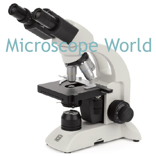
Plastic or Metal: Only those microscopes made of die-cast metal are sturdy enough to withstand viewing without vibration interference. Plastic microscopes are lighter, but they are also less stable and easily damaged. The lenses (optics) in the microscope should always be made of glass. Plastic optics offer a blurry image and can be frustrating to use, especially for children.

Microscope Magnification: When using a biological (high power) microscope, magnification above 1000x is frowned upon, as it offers empty magnification that will not be clear and crisp.

Monocular, Binocular or Trinocular Microscope: A monocular microscope has a single eyepiece for viewing. This is perfect for occasional viewing through the microscope. Binocular microscopes offer two eyepieces and are more comfortable for viewing items over a longer period of time. Trinocular microscopes provide two eyepieces for viewing the specimen, along with an additional eyepiece for attaching a camera (the trinocular port).

Monocular Microscope
Binocular Microscope

Trinocular Microscope
Illumination: The most common microscope illuminators offered include tungsten, fluorescent, halogen and LED. Tungsten and halogen illuminators heat up, so if viewing live specimens that could be damaged by heat, it is recommended to avoid these lights. Halogen and LED illuminators are the brightest and of all the light microscope options, LED is usually preferable because of its cool, long-lasting light bulbs.

Focusing: Most microscopes will offer either only coarse focusing (single focusing knob) or coarse and fine focusing (two focusing knobs). Coarse and fine focusing is generally required for clear viewing of any specimens above 40x magnification.

X-Y Stage: A microscope mechanical stage allows the user to move the stage from left to right or forward and backward by using one of two knobs. Slight adjustments can be made in the stage position, making it easier to keep the specimen in the field of view while looking at it.
Viewing all 422 articles
Browse latest View live